. 2019 Apr 8;219(8):1264-1273.
doi: 10.1093/infdis/jiy635.

Human mAbs to Staphylococcus aureus IsdA Provide Protection Through Both Heme-Blocking and Fc-Mediated Mechanisms

Affiliations

Human mAbs to Staphylococcus aureus IsdA Provide Protection Through Both Heme-Blocking and Fc-Mediated Mechanisms

Monique R Bennett et al. J Infect Dis. .

Abstract

The nutrient metal iron plays a key role in the survival of microorganisms. The iron-regulated surface determinant (Isd) system scavenges heme-iron from the human host, enabling acquisition of iron in iron-deplete conditions in Staphylococcus aureus during infection. The cell surface receptors IsdB and IsdH bind hemoproteins and transfer heme to IsdA, the final surface protein before heme-iron is transported through the peptidoglycan. To define the human B-cell response to IsdA, we isolated human monoclonal antibodies (mAbs) specific to the surface Isd proteins and determined their mechanism of action. We describe the first isolation of fully human IsdA and IsdH mAbs, as well as cross-reactive Isd mAbs. Two of the identified IsdA mAbs worked in a murine septic model of infection to reduce bacterial burden during staphylococcal infection. Their protection was a result of both heme-blocking and Fc-mediated effector functions, underscoring the importance of targeting S. aureus using diverse mechanisms.

Keywords: Staphyloccocus aureus; IsdA; NEAr iron transporter; antibodies; human; iron-regulated surface determinant system.


Figures

Figure 1.
Figure 1.
Competition-binding of IsdA- or IsdB-specific antibodies. Biolayer interferometry was used to perform competition-binding studies on the Isd-specific antibody panel. Either IsdA (left panel) or IsdB (right panel) was biotinylated and loaded onto streptavidin tips before testing of binding to a first and then second antibody. The percent binding of the second antibody to the biosensor tip in the presence of bound first antibody. Black boxes indicate the first antibody blocked binding of the second antibody by more than 70%. Grey boxes indicate the first antibody partially blocked binding of the second antibody, with 31%–75% residual binding of the second remaining. White boxes indicate the first antibody did not block binding of the second antibody to the biosensor tip. Assigned competition-binding groups are outlined in color. Certain cross-competing antibodies did not bind well to both IsdA and IsdB using biolayer interferometry, suggesting that they may be members of an additional competition-binding group that could not be tested in this way. Individual antibodies were competed in at least 2 independent experiments.
Figure 2.
Figure 2.
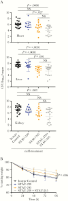
Isd-specific antibodies reduce bacterial burden in a systemic murine model of infection. A, Seven-week-old female BALB/c mice were inoculated retro-orbitally with an OD of 0.4 suspension (approximately107 colony forming units [CFU]) S. aureus strain Newman. After 96 hours, the heart, livers, and kidneys of the infected mice were harvested in cold phosphate-buffered saline. CFU counts were determined by serial dilution plating on TSA. P values were determined by a Kruskal-Wallis nonparametric test and the Dunn Procedure for multiple comparisons. Data are expressed as the geometric mean ± geometric standard deviation. B, Weight loss over the course of 96 hours was calculated as a percentage of the starting weight. P values were determined by ANOVA. Experiments were performed 3 independent times and the pooled data are shown.
Figure 3.
Figure 3.
Partial inhibition of Staphylococcus aureus hemoglobin-dependent growth. S. aureus strain Newman was cultured overnight in RPMI with 1 μM ethylenediamine-N,N′-bis(2-hydroxyphenylacetic acid) and normalized to OD600 1 before being subcultured and grown in broth for 34 hours. Statistically significant differences were determined by a Mann–Whitney test, and P values are shown where the hemoglobin-treated group was set as the comparator. Data shown are representative of 3 independent experiments.
Figure 4.
Figure 4.
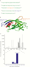
Binding sites and conservation of STAU-239 and STAU-245 on iron-regulated surface determinant A-N1 (IsdA-N1). Hydrogen-deuterium exchange mass spectrometry (HDX-MS) was used to map the binding sites of STAU-239 and STAU-245 onto IsdA. A, The sequence of IsdA-N1 is shown and amino acids with the strongest decrease in deuterium uptake in the presence of STAU-239 (blue) or STAU-245 (orange) and IsdA are colored. B, The structure of the crystallized NEAr iron transporter (NEAT) domain of IsdA (Research Collaboratory for Structural Bioinformatics Protein Data Bank Identification [PDB ID]: 2ITF) is shown binding heme (red) with the predicted binding sites of STAU-239 (blue) and STAU-245 (orange). C, Amino acid changes in the S. aureus strain Newman IsdA protein were determined from 42522 S. aureus genomes. Frequency distribution of all amino acid changes across the 350 residues of IsdA in 42522 strains is shown (top). Amino acid changes observed specifically in the STAU-239 binding site (amino acid positions 159 and 167) are highlighted (bottom).
Figure 5.
Figure 5.
Monoclonal antibody (mAb) STAU-239 + STAU-245 Fc mutants do not reduce bacterial burden in systemic murine model of infection. A, Seven-week-old female BALB/c mice were inoculated retro-orbitally with OD600 0.4 suspension of Staphylococcus aureus strain Newman. Mice were given 1 of 2 mixes: (STAU-239 + STAU-245 IgG) or (STAU-239 + STAU-245 LALA [L234A, L235A] variant) by the intraperitoneal route 2 hours before infection. After 96 hours, the heart, livers, and kidneys of the infected mice were harvested in cold phosphate-buffered saline. Colony forming units (CFU) were determined by serial dilution plating on TSA. P values were determined by a Kruskal-Wallis nonparametric test and the Dunn Procedure for multiple comparisons. Data are expressed as the geometric mean ± geometric standard deviation. B, Weight loss over the 96-hour infection was calculated as a percentage of the starting weight. P values were determined by ANOVA. Experiments were performed 2 independent times and the pooled data are shown.

References

    1. Hood MI, Skaar EP. Nutritional immunity: transition metals at the pathogen-host interface. Nat Rev Microbiol 2012; 10:525–37. - PMC - PubMed
    1. Skaar EP. The battle for iron between bacterial pathogens and their vertebrate hosts. PLoS Pathog 2010; 6:e1000949. - PMC - PubMed
    1. Skaar EP, Humayun M, Bae T, DeBord KL, Schneewind O. Iron-source preference of Staphylococcus aureus infections. Science 2004; 305:1626–8. - PubMed
    1. Mazmanian SK, Skaar EP, Gaspar AH, et al. . Passage of heme-iron across the envelope of Staphylococcus aureus. Science 2003; 299:906–9. - PubMed
    1. Grigg JC, Vermeiren CL, Heinrichs DE, Murphy ME. Haem recognition by a Staphylococcus aureus NEAT domain. Mol Microbiol 2007; 63:139–49. - PubMed

Publication types

Substances