. 2019 Jan 10:293:158-171.
doi: 10.1016/j.jconrel.2018.11.025. Epub 2018 Nov 27.

Oral co-administration of a bacterial protease inhibitor in the vaccine formulation increases antigen delivery at the intestinal epithelial barrier

Affiliations

Oral co-administration of a bacterial protease inhibitor in the vaccine formulation increases antigen delivery at the intestinal epithelial barrier

Lorena M Coria et al. J Control Release. .

Abstract

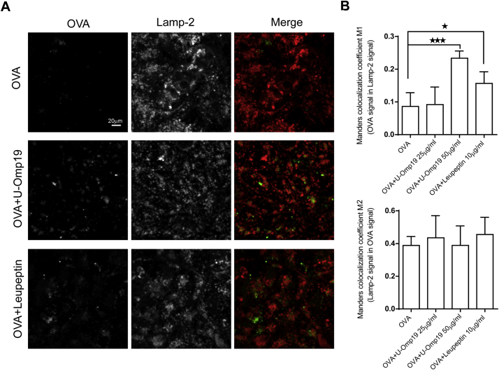
The study of capture and processing of antigens (Ags) by intestinal epithelial cells is very important for development of new oral administration systems. Efficient oral Ag delivery systems must resist enzymatic degradation by gastric and intestinal proteases and deliver the Ag across biological barriers. The recombinant unlipidated outer membrane protein from Brucella spp. (U-Omp19) is a protease inhibitor with immunostimulatory properties used as adjuvant in oral vaccine formulations. In the present work we further characterized its mechanism of action and studied the interaction and effect of U-Omp19 on the intestinal epithelium. We found that U-Omp19 inhibited protease activity from murine intestinal brush-border membranes and cysteine proteases from human intestinal epithelial cells (IECs) promoting co-administered Ag accumulation within lysosomal compartments of IECs. In addition, we have shown that co-administration of U-Omp19 facilitated the transcellular passage of Ag through epithelial cell monolayers in vitro and in vivo while did not affect epithelial cell barrier permeability. Finally, oral co-delivery of U-Omp19 in mice induced the production of Ag-specific IgA in feces and the increment of CD103+ CD11b- CD8α+ dendritic cells subset at Peyer's patches. Taken together, these data describe a new mechanism of action of a mucosal adjuvant and support the use of this rationale/strategy in new oral delivery systems for vaccines.

Keywords: Bacterial protease inhibitor; Enterocytes; Intestinal barrier; Oral delivery; Vaccine adjuvant.


Figures

Unlabelled Image
Graphical abstract
Fig. 1
Fig. 1
U-Omp19 partially inhibits brush border and intracellular proteases from enterocytes. A. Brush-border membranes (50 μg) proteolytic activity was determined after incubation for 1 h with different amounts of U-Omp19 (25, 50 and 100 μg/ml) by adding Casein-BODIPY substrate. BSA was used as negative control. Data are presented as percentage of casein digestion ± SEM. *p < 0.05; **p < 0.01; ***p < 0.001 vs. no protease inhibitor (--). ANOVA followed by Bonferroni multiple comparison test B. U-Omp19 increases Ag amount inside human intestinal epithelial cells. Caco-2 and HT29 epithelial cell lines were incubated with OVA-FITC alone or plus U-Omp19 during 3 h and then MFI was determined by flow cytometry. Data are presented as histograms and bar graphs ± SEM. C. U-Omp19 inhibits Ag digestion inside human epithelial cells. Caco-2 and HT29 epithelial cell lines were incubated with DQ-OVA or Casein-BODIPY alone or plus U-Omp19 during 3 h and the fluorescence measured by flow cytometry. Results are presented as histograms and bar graphs (mean fluorescence intensity -MFI- ± SEM) for each group. *p < 0.05; **p < 0.01; ***p < 0.001 vs. Ag alone. (OVA-FITC, DQ-OVA or Casein-BODIPY). t-test.
Fig. 2
Fig. 2
U-Omp19 inhibits cathepsin L activity in human intestinal epithelial cells. Caco-2 (A) and HT29 (B) cells were incubated during 1 h with different concentrations of U-Omp19 or Leupeptin. Then, a specific fluorescence quenched substrate for cathepsin L was added and the reaction was measured in a microplate fluorescence reader for 4 h. Data are shown as arbitrary fluorescence units or percentage of remaining cathepsin L activity over time. C. Confocal microscopy analysis of Caco-2 cells after incubation with the specific quenched substrate for cathepsin L (green) and Lysotracker (red) in presence of medium, U-Omp19 or Leupeptin. Images are representative of most cells examined by confocal microscopy. Colocalization was analyzed by Manders colocalization coefficient (M1 and M2). Data are means of Manders coefficient ± SEM. Scale bars 5 μm. *p < 0.05; **p < 0.01; ***p < 0.001 vs. medium. ANOVA followed by Bonferroni multiple comparison test. (For interpretation of the references to colour in this figure legend, the reader is referred to the web version of this article.)
Fig. 3
Fig. 3
U-Omp19 co-delivery induces antigen accumulation at enterocytes lysosomes. Confocal scanning microscopy analysis of Caco-2 cells treated with OVA–Alexa Fluor 647 (25 μg/ml) alone or plus U-Omp19 (25 or 50 μg/ml) and Leupeptin (10 μg/ml). After 3 h of incubation cells were fixed, permeabilized, and stained with mAb anti–human Lamp-2. Anti-mouse IgG coupled to Alexa Fluor 546 (red) was used as secondary Ab. Images are representative of most cells examined by confocal microscopy. The merge between Lamp-2/OVA is shown. Quantification of colocalization OVA/Lamp-2 (M1) and Lamp-2/OVA (M2) was analyzed by Manders overlap coefficient. Data are means of Manders coefficients ± SEM present in 4–5 images of each condition. Scale bars 20 μm. * p < 0.05; ***p < 0.001 vs OVA. ANOVA followed by Bonferroni multiple comparison test. (For interpretation of the references to colour in this figure legend, the reader is referred to the web version of this article.)
Fig. 4
Fig. 4
U-Omp19 facilitates Ag transport through epithelial cell monolayers. Transwell plates cultured Caco-2 cell monolayers were incubated with OVA-Alexa Fluor 647 alone (0.5 mg/ml) or in presence of U-Omp19 or Leupeptin during 3 h. Then, fluorescence in cell lysates (A) and basolateral medium (B) were measured in a microplate fluorescence reader. Result were pooled from 3 different experiments and data are means of percentage of OVA ± SEM. *p < 0.05; **p < 0.01 vs OVA. ANOVA followed by Bonferroni multiple comparison test. Western blot analysis and quantitative densitometry of OVA signal was performed in precipitated proteins of cell lysates (C) and basolateral medium (D). Calnexin was used as internal loading control in the case of cell lysates. Data are presented as means of relative density of OVA ± SD. *p < 0.05 vs OVA. ANOVA followed by Bonferroni multiple comparison test.
Fig. 5
Fig. 5
Ag fate after oral co-delivery with U-Omp19 in mice intestine sections. BALB/c mice were orally co-delivered with OVA-Alexa Fluor 647 (white) + U-Omp19 and 2 h later intestines were excised. Intestine cryosections were stained with UEA (green) and WGA staining (red) for epithelial cells. Yellow arrows indicate OVA-Alexa Fluor 647 (white) at duodenum (A), jejunum (B) and ileum (C). Images are representative of two independent experiments. Scale bars 100 μm (left panels, 10× objective) or 20 μm (inset panels). D. Jejunum and Ileum sections were labeled with rabbit anti-U-Omp19 polyclonal Ab or mouse anti-CD11c mAb as primary antibodies and then anti-rabbit Alexa Fluor 488 (green) or anti-mouse Alexa Fluor 546 (red) secondary antibodies. Images shown OVA-AF647 (white), U-Omp19 (green) or CD11c (red) signal separately and merge of 3 signals and DIC (right panels). Scale bars 50 μm. (For interpretation of the references to colour in this figure legend, the reader is referred to the web version of this article.)
Fig. 6
Fig. 6
U-Omp19 increases dendritic cells subsets at Peyer's patches after oral delivery. BALB/c mice were orally administered with OVA alone or plus U-Omp19 and 18 h later DC populations were analyzed in Peyer's patches. Flow cytometry analysis of different DC populations were performed using anti-CD11c, anti-MHCII, anti-CD11b, anti-CD103, and anti-CD8α antibodies. Gating strategy is shown in A. Results are presented as percentage of total cells and in mean numbers of cells obtained for each mouse ± SEM and are representative of 3 different experiments. *p < 0.05, **p < 0.01 vs. OVA group. Mann Whitney U test.
Fig. 7
Fig. 7
U-Omp19 in the oral vaccine formulation increases the frequency of mucosal DCs bearing the co-delivered Ag. BALB/c mice were orally administered with OVA Alexa Fluor 647 alone or plus U-Omp19 and 4 h later DC populations were analyzed in MLNs. Flow cytometry analysis of different DC populations were performed using anti-CD11c, anti-MHCII, anti-CD11b, anti-CD103, and anti-CD8α antibodies. Gating strategy is shown in A. Results are presented as percentage of OVA-AF647+ cells in the different populations of DCs (CD11c+ MHCII+) ± SEM. *p < 0.05 vs. OVA group. Mann Whitney U test.
Fig. 8
Fig. 8
U-Omp19 increases CTB half-life and immunogenicity after oral co-delivery. A. Caco-2 epithelial cell line was incubated with CTB-Alexa Fluor 647 (10 μg/ml) alone or plus U-Omp19 (50 μg/ml) or Leupeptin (10 μg/ml) during 3 h and then MFI was determined by flow cytometry. Data are presented as histograms and bar graphs ± SEM. B. U-Omp19 co-delivery with CTB increases CTB accumulation inside the lysosome of intestinal epithelial cells. Confocal microscopy analysis of Caco-2 cells treated with CTB-Alexa Fluor 647 (10 μg/ml, green) alone or plus U-Omp19 (25 or 50 μg/ml) and Leupeptin (10 μg/ml). After 3 h of incubation cells were fixed, permeabilized, and stained with mAb anti–human Lamp-2. Anti-mouse IgG coupled to Alexa Fluor 546 (red) was used as secondary Ab. Images are representative of most cells examined by confocal microscopy. C. Quantification of colocalization was analyzed by Manders overlap coefficient. Data are means of Manders coefficients ± SEM present in 4–5 images of each condition. *p < 0.05; **p < 0.01 vs CTB alone. ANOVA followed by Bonferroni multiple comparison test. D-E. U-Omp19 induces specific antibody responses after oral co-delivery with CTB. BALB/c mice (n = 6/group) were orally immunized on days 1, 2, 3, 8, 9 and 10 with saline, CTB (10 μg) or CTB plus U-Omp19 (150 μg). Specific IgA and IgG levels anti-CTB and anti-U-Omp19 were evaluated in feces and sera by ELISA. Anti-CTB IgG1 and IgG2a levels were evaluated in sera. Data are means of OD 450 ± SEM for IgA responses and as IgG titers or titer ratio ± SEM for anti-CTB IgG, IgG1 or IgG2a. *p < 0.05 vs CTB group. Mann Whitney U test. (For interpretation of the references to colour in this figure legend, the reader is referred to the web version of this article.)

References

    1. Pasetti M.F., Simon J.K., Sztein M.B., Levine M.M. Immunology of gut mucosal vaccines. Immunol. Rev. 2011;239:125–148. - PMC - PubMed
    1. Kew O., Morris-Glasgow V., Landaverde M., Burns C., Shaw J., Garib Z., Andre J., Blackman E., Freeman C.J., Jorba J., Sutter R., Tambini G., Venczel L., Pedreira C., Laender F., Shimizu H., Yoneyama T., Miyamura T., van Der Avoort H., Oberste M.S., Kilpatrick D., Cochi S., Pallansch M., de Quadros C. Outbreak of poliomyelitis in Hispaniola associated with circulating type 1 vaccine-derived poliovirus. Science. 2002;296:356–359. - PubMed
    1. Lycke N. Recent progress in mucosal vaccine development: potential and limitations. Nat. Rev. Immunol. 2012;12:592–605. - PubMed
    1. Bernkop-Schnürch A. Springer; Dordrecht; New York: 2009. Oral Delivery of Macromolecular Drugs: Barriers, Strategies, and Future Trends.
    1. Mahato R.I., Narang A.S., Thoma L., Miller D.D. Emerging trends in oral delivery of peptide and protein drugs. Crit. Rev. Ther. Drug Carrier Syst. 2003;20:153–214. - PubMed

Publication types

MeSH terms