Case Reports
. 2019 Apr:57:313-316.
doi: 10.1016/j.mri.2018.11.016. Epub 2018 Nov 27.

7-Tesla MRI of the brain in a research subject with bilateral, total knee replacement implants: Case report and proposed safety guidelines

Affiliations
Case Reports

7-Tesla MRI of the brain in a research subject with bilateral, total knee replacement implants: Case report and proposed safety guidelines

Giuseppe Barisano et al. Magn Reson Imaging. 2019 Apr.

Abstract

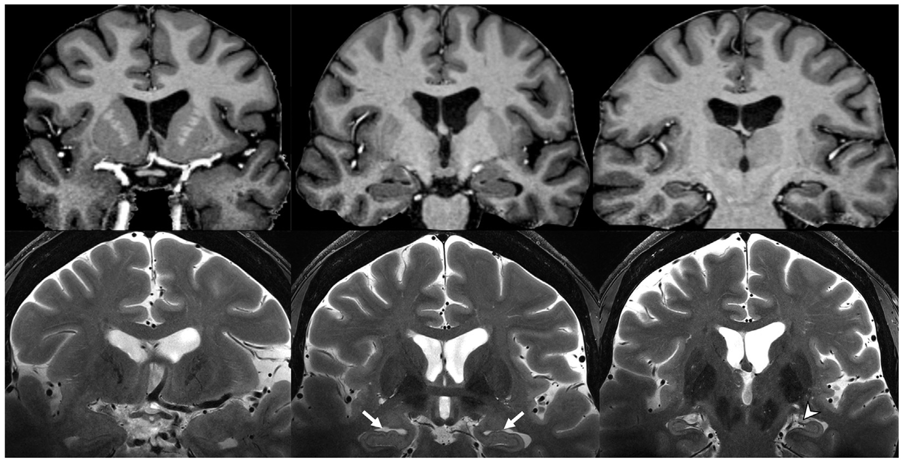
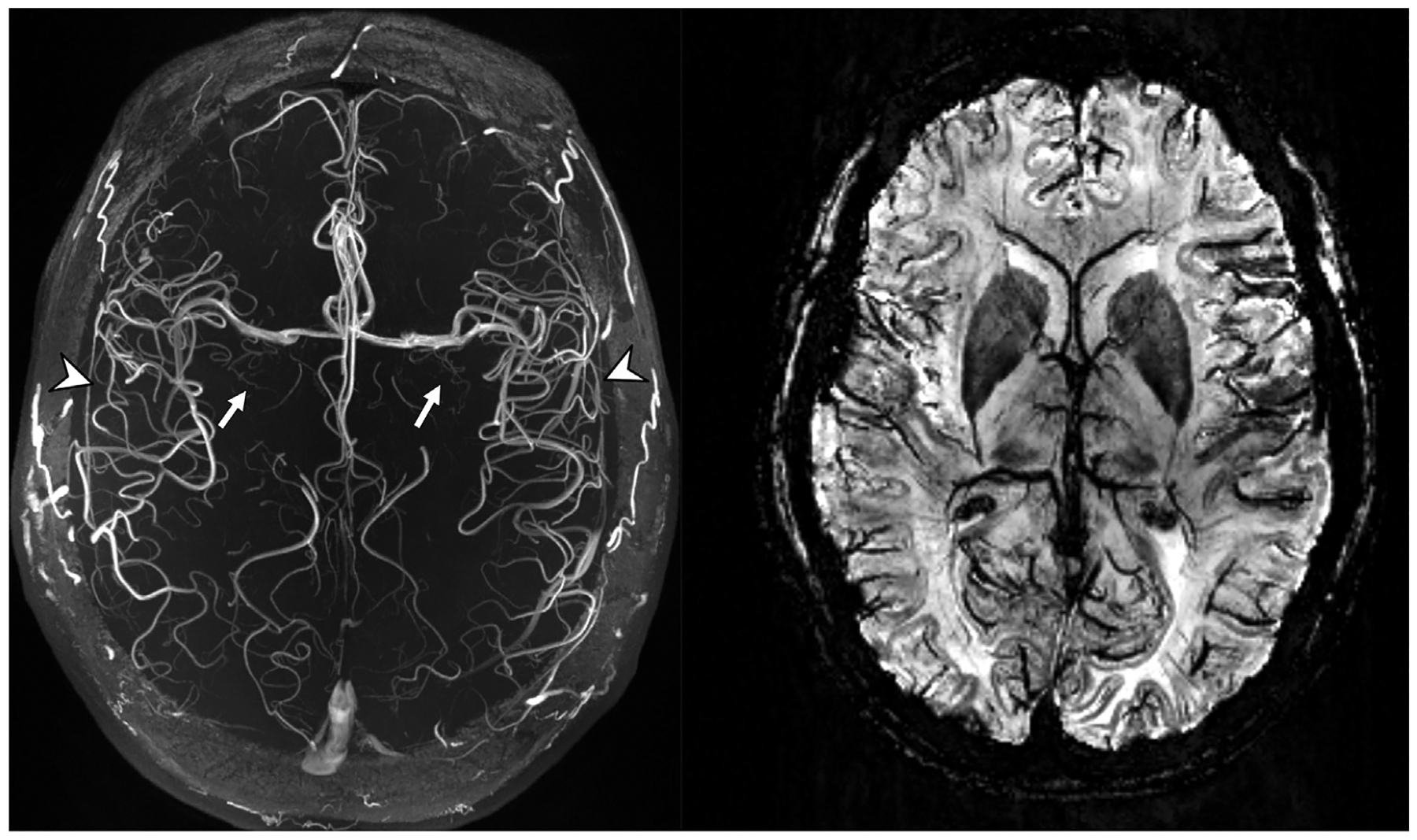
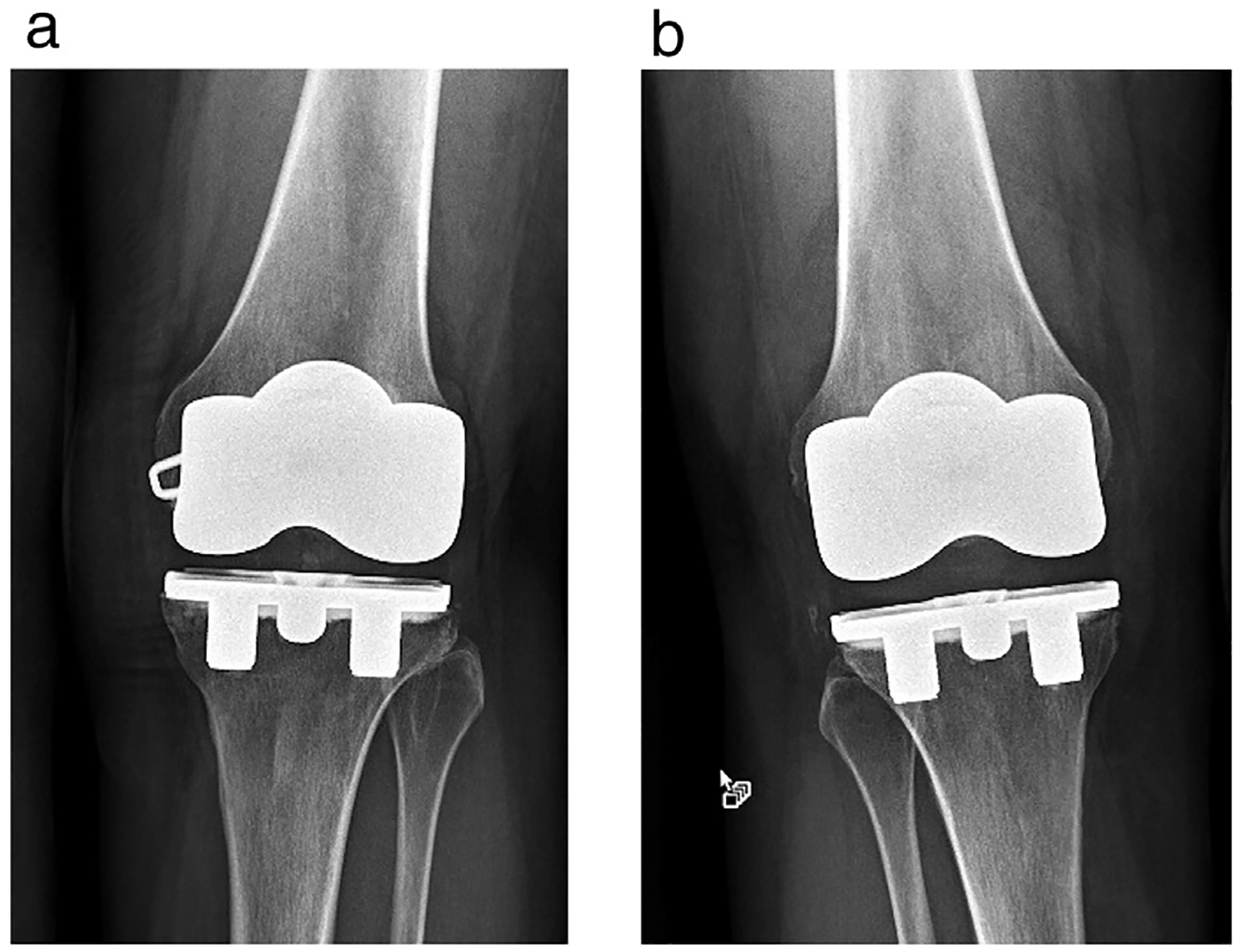
Recently, the first 7-T MR system was approved for clinical use in the United States. Unfortunately, relatively few metallic implants have undergone testing to determine if they are acceptable or pose hazards to research subjects and patients at this ultra-high-field strength. Therefore, in lieu of not performing a research or clinical MRI exam at 7-T, the supervising physician may make a decision to scan the individual with an untested metallic implant based on an analysis of the risks vs. the benefits. We present a case report of a research subject with bilateral, total knee replacement implants that safely underwent MRI of the brain at 7-T and provide guidelines for healthcare professionals to follow in order to ensure safety in research subjects or patients with metallic implants referred for 7-T scans.

Keywords: 7-Tesla; Brain; MRI implants; MRI safety; Magnetic resonance imaging (MRI).


Figures

Fig. 1.
Fig. 1.
Skull-stripped coronal plane T1 MP2RAGE (top row) and unprocessed T2-weighted (bottom row) MR images. Note the contrast between grey and white matter on the T1 MP2RAGE, and the hippocampal digitations (white arrows), fimbria (arrowhead), and stratum layers of the hippocampus resolved on the T2-weighted images at 7-T. (MP2RAGE, magnetization prepared, two rapid-acquisition gradient-echo).
Fig. 2.
Fig. 2.
Axial plane TOF-MRA MIP (left) and SWI MinIP (right) MR images. Note the conspicuity of the lenticulostriate arteries (white arrows) arising from the MCAs, the high contrast seen in the insular branches (arrowheads) of the MCAs, and the high susceptibility and spatial resolution allowing better definition of deoxyhemoglobin in the smaller veins on the SWI image. (MCA, middle cerebral artery; MinIP, minimum intensity projection; MIP, maximum intensity projection; SWI, susceptibility-weighted imaging; TOF-MRA, time-of-flight, magnetic resonance angio-graphy).
Fig. 3.
Fig. 3.
X-rays showing the TKR implants for the research subject that underwent MRI of the brain at 7-T. (a) Left knee, (b) Right knee.

References

    1. U.S. Department of Health and Human Services, U.S. Food and Drug Administration. FDA clears first 7 T magnetic resonance imaging device. https://www.fda.gov/NewsEvents/Newsroom/PressAnnouncements/ucm580154.htm, Accessed date: 12 October 2017.
    1. Trattnig S, Springer E, Bogner W, et al. Key clinical benefits of neuroimaging at 7 T. NeuroImage 2018;168:477–89. - PMC - PubMed
    1. Balchandani P, Naidich TP. Ultra-high-field MR neuroimaging. AJNR Am J Neuroradiol 2015;36:1204–15. - PMC - PubMed
    1. Barisano G, Sepehrband F, Ma S, et al. Clinical 7T MRI: Are we there yet? A review about magnetic resonance imaging at ultra-high field. Br J Radiol. 2018;91:20180492. - PMC - PubMed
    1. Kraff O, Quick HH. 7 T: physics, safety, and potential clinical applications. J Magn Reson Imaging 2017;46:1573–89. 10.1002/jmri.25723. - DOI - PubMed

Publication types