Review
. 2019 Jul 1;1865(7):1831-1844.
doi: 10.1016/j.bbadis.2018.11.010. Epub 2018 Nov 27.

As time flies by: Investigating cardiac aging in the short-lived Drosophila model

Affiliations
Review

As time flies by: Investigating cardiac aging in the short-lived Drosophila model

Anna C Blice-Baum et al. Biochim Biophys Acta Mol Basis Dis. .

Abstract

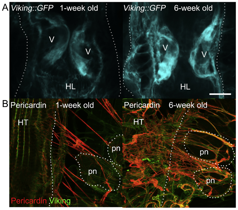
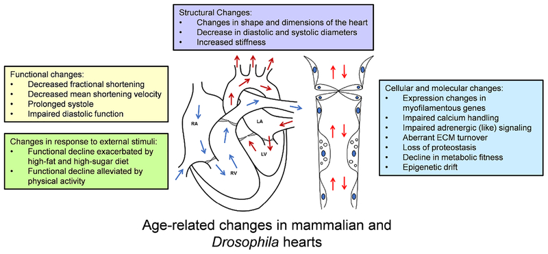
Aging is associated with a decline in heart function across the tissue, cellular, and molecular levels. The risk of cardiovascular disease grows significantly over time, and as developed countries continue to see an increase in lifespan, the cost of cardiovascular healthcare for the elderly will undoubtedly rise. The molecular basis for cardiac function deterioration with age is multifaceted and not entirely clear, and there is a limit to what investigations can be performed on human subjects or mammalian models. Drosophila melanogaster has emerged as a useful model organism for studying aging in a short timeframe, benefitting from a suite of molecular and genetic tools and displaying highly conserved traits of cardiac senescence. Here, we discuss recent advances in our understanding of cardiac aging and how the fruit fly has aided in these developments.

Keywords: Cardiac aging; Epigenetics; Fruit fly; Obesity; Proteostasis.


Figures

Figure 1:
Figure 1:. The adult Drosophila heart tube and associated structures.
(A) The abdominally located adult fruit fly heart tube (HT) contains roughly 80 cardiomyocytes, arranged along two opposing bilateral rows. These aligned cells form a central luminal space and are flanked by adjacent pericardial nephrocytes (pns) that filter the hemolymph (insect’s blood) and alary muscles (ams), which tether the heart to the dorsal cuticle [, –258]. The alary muscles span from epidermal attachments to the heart and contact the heart tube indirectly by an interface formed from extracellular matrix components, such as the collagens Pericardin and Viking. They serve as a flexible heart suspension system to maintain the cardiac tube in an anatomically correct position. (B) Heart tubes showing cardiac collagen-IV (Viking) and Pericardin (a collagen IV-like protein) deposition. Hemolymph enters the heart through the ostia (os), which are inflow openings formed by specialized cardiomyocytes. The adult Drosophila heart has three intracardiac valves (v), which subdivide the organ into distinct chambers, close the luminal space during systole, and permit unidirectional flow. Pericardial nephrocytes are analogous to mammalian reticuloendothelial cells and, being situated close to ostia, are ideally placed to filter the passing hemolymph entering the heart tube. Scale bar = 100μm. (C) Illustration depicting the aforementioned anatomical structures.
Figure 2.
Figure 2.. Changes to cardiac collagen deposition in aging fly hearts.
Under normal laboratory conditions, the fly’s lifespan is approximately 8–10 weeks. Prior to the onset of mortality, in the general population at around six weeks, a significant increase in the amount of cardiac collagen-IV (Viking) and Pericardin (a collagen IV-like protein, restricted to the heart) can be seen around the heart. With advanced age, there is an increased accumulation of collagen. Confocal micrographs demonstrate age-associated changes to (A) Viking::GFP levels on the luminal side of the valve cells and (B) the Pericardin network. Scale bar = 20μm.
Figure 3:
Figure 3:. Hallmarks of cardiac aging are shared across species.
The changes reported above are well-conserved, highly interdependent, and contribute to a decline in overall cardiac function over time. Note that some changes may arise in response to others and should be studied in more depth. RA = right atrium, RV = right ventricle, LA = left atrium, and LV = left ventricle. Arrows indicate flow direction.

References

    1. López-Otín C, Blasco MA, Partridge L, et al. (2013) The hallmarks of aging. Cell 153 - PMC - PubMed
    1. Go AS, Mozaffarian D, Roger VL, et al. (2014) Heart disease and stroke statistics−-2014 update: a report from the American Heart Association. Circulation 129:e28–e292. doi: 10.1161/01.cir.0000441139.02102.80 - DOI - PMC - PubMed
    1. Barquera S, Pedroza-Tobías A, Medina C, et al. (2015) Global overview of the epidemiology of atherosclerotic cardiovascular disease. Arch. Med. Res 46:328–338 - PubMed
    1. Heidenreich PA, Trogdon JG, Khavjou OA, et al. (2011) Forecasting the future of cardiovascular disease in the United States: A policy statement from the American Heart Association. Circulation 123:933–944. doi: 10.1161/CIR.0b013e31820a55f5 - DOI - PubMed
    1. Schmid T (2015) Costs of treating cardiovascular events in Germany: a systematic literature review. Health Econ. Rev 5 - PMC - PubMed

Publication types