. 2018 Nov 30;19(1):426.
doi: 10.1186/s12891-018-2353-7.

Tendinosis-like changes in denervated rat Achilles tendon

Affiliations

Tendinosis-like changes in denervated rat Achilles tendon

Roine El-Habta et al. BMC Musculoskelet Disord. .

Abstract

Background: Tendon disorders are common and lead to significant disability and pain. Our knowledge of the 'tennis elbow', the 'jumpers knee', and Achilles tendinosis has increased over the years, but changes in denervated tendons is yet to be described in detail. The aim of the present study was to investigate the morphological and biochemical changes in tendon tissue following two weeks of denervation using a unilateral sciatic nerve transection model in rat Achilles tendons.

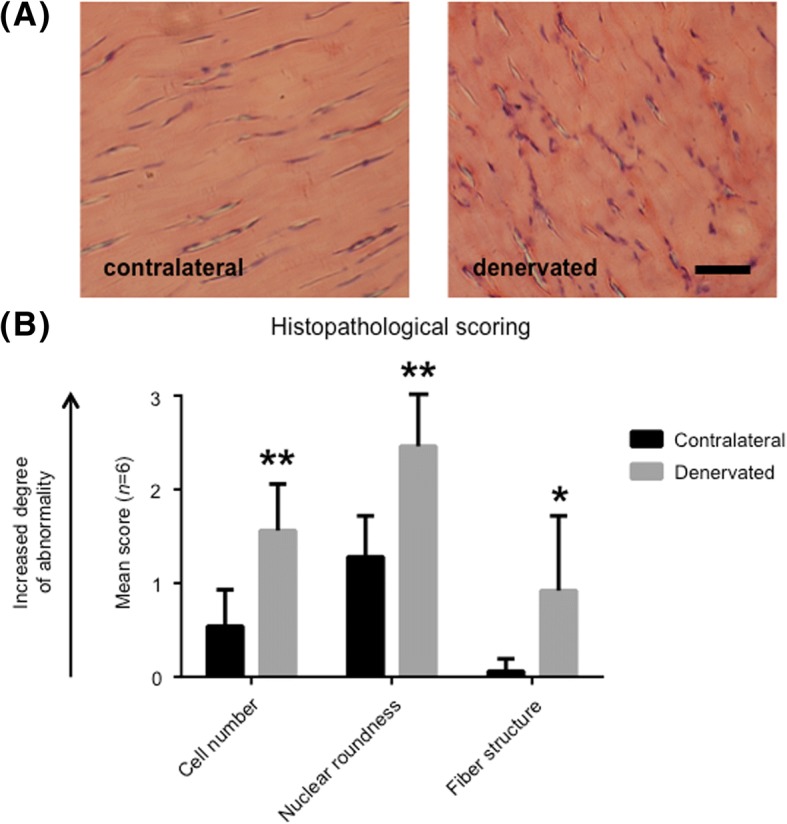
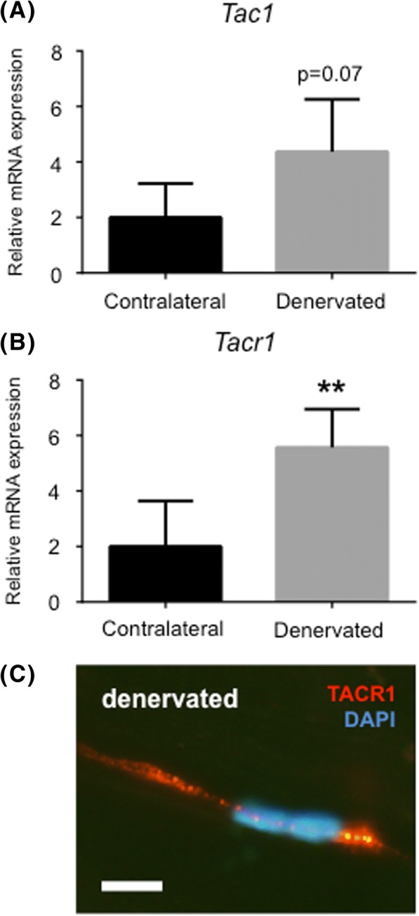
Methods: Tendons were compared with respect to cell number, nuclear roundness, and fiber structure. The non-denervated contralateral tendon served as a control. Also, the expression of neuromodulators such as substance P and its preferred receptor neurokinin-1 receptor, NK-1R, was evaluated using real-time qRT-PCR.

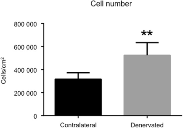
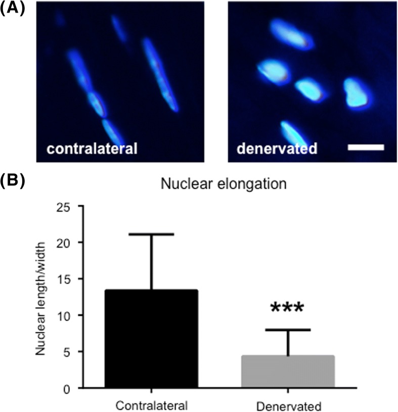
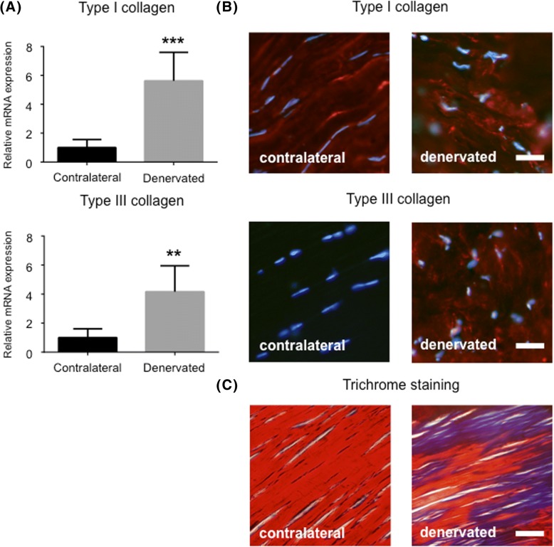
Results: Our results showed that denervated tendons expressed morphological changes such as hypercellularity; disfigured cells; disorganization of the collagen network; increased production of type III collagen; and increased expression of NK-1R.

Conclusion: Taken together these data provide new insights into the histopathology of denervated tendons showing that denervation causes somewhat similar changes in the Achilles tendon as does tendinosis in rats.

Keywords: Collagen; Denervation; Rat; Substance P; Tendinosis.


Conflict of interest statement

Ethics approval and consent to participate

The animal care and experimental procedures were carried out in accordance with the Directive 2010/63/EU of the European Parliament and of the Council on the protection of animals used for scientific purposes. The study was also approved by the Northern Swedish Committee for Ethics in Animal Experiments (No. A186–12).

Consent for publication

Not applicable.

Competing interests

The authors declare that they have no competing interests.

Publisher’s Note

Springer Nature remains neutral with regard to jurisdictional claims in published maps and institutional affiliations.

Figures

Fig. 1
Fig. 1
Histology of denervated rat Achilles tendon. (a) Hematoxylin-eosin staining of the middle part of the Achilles tendon two weeks after unilateral sciatic nerve transection. The rats were allowed to move freely post-surgery. Both sides, i.e. denervated and contralateral, are shown. Bar = 20 μm. (b) Histological sections were examined in a light microscope and given a score from 0 to 3 (0 = normal, and 3 = pathological). All examined parameters were significantly increased in the denervated group compared to the contralateral healthy leg. * indicates p < 0.05, ** indicates p < 0.01. Values of p < 0.05 were considered statistically significant
Fig. 2
Fig. 2
Pronounced increase in the number of cells in denervated tendons. Mean cell number from six independent animals. Denervated side compared to the contralateral side. Overall, there was a significant increase (approximately 70%) in the number of cells in denervated tendons compared to contralateral tendons. ** indicates p < 0.01. Values of p < 0.05 were considered statistically significant
Fig. 3
Fig. 3
Increased nuclear roundness in denervated tendons. (a) Nuclei stained with DAPI revealing increased nuclear roundness in denervated samples compared to contralateral samples. Bar = 5 μm. (b) Ratio between nuclear length and width, measured with ImageJ. The lower the value, the greater the nuclear roundness is. *** indicates p < 0.001. Values of p < 0.05 were considered statistically significant
Fig. 4
Fig. 4
Disturbed production and organization of collagen in denervated tendons. (a) Increased mRNA expression of type I and type III collagen in denervated tendons. (b) Expression of collagen I and III protein (stained red) in contralateral (left column) and denervated (right column) tendons. DAPI was used to stain the nuclei (blue). Bar = 20 μm. (c) Trichrome stain revealing disorganized collagen (blue) in denervated tendons. Bar = 20 μm. ** indicates p < 0.01, *** indicates p < 0.001. Values of p < 0.05 were considered statistically significant
Fig. 5
Fig. 5
Denervated tendons express molecules associated with tendinosis. (a) Expression of genes related to tendinosis using qRT-PCR: substance P (Tac1) and its preferred receptor NK-1R (Tacr1). (b) Immunohistochemistry showing positive staining for NK-1R in a denervated tendon. Bar = 5 μm. ** indicates p < 0.01. Values of p < 0.05 were considered statistically significant

References

    1. Kjaer M. Role of extracellular matrix in adaptation of tendon and skeletal muscle to mechanical loading. Physiol Rev. 2004;84(2):649–698. doi: 10.1152/physrev.00031.2003. - DOI - PubMed
    1. Kannus P, Paavola M, Paakkala T, Parkkari J, Jarvinen T, Jarvinen M. Pathophysiology of overuse tendon injury. Radiologe. 2002;42(10):766–770. doi: 10.1007/s00117-002-0795-1. - DOI - PubMed
    1. Jozsa L, Kannus P. Histopathological findings in spontaneous tendon ruptures. Scand J Med Sci Sports. 1997;7(2):113–118. doi: 10.1111/j.1600-0838.1997.tb00127.x. - DOI - PubMed
    1. Holm C, Kjaer M, Eliasson P. Achilles tendon rupture--treatment and complications: a systematic review. Scand J Med Sci Sports. 2015;25(1):e1–10. doi: 10.1111/sms.12209. - DOI - PubMed
    1. Thakkar RS, Thakkar SC, Srikumaran U, McFarland EG, Fayad LM. Complications of rotator cuff surgery-the role of post-operative imaging in patient care. Br J Radiol. 2014;87(1039):20130630. doi: 10.1259/bjr.20130630. - DOI - PMC - PubMed