. 2018 Nov 29;51(1):53.
doi: 10.1186/s40659-018-0201-8.

The deubiquitinase USP38 affects cellular functions through interacting with LSD1

Affiliations

The deubiquitinase USP38 affects cellular functions through interacting with LSD1

Wenbin Liu et al. Biol Res. .

Abstract

Background: Deubiquitination is a posttranslational protein modification prevalent in mammalian cells. Deubiquitinases regulate the functions of the target protein by removing its ubiquitin chain. In this study, the effects of the deubiquitinase USP38's functions on the LSD1 protein and on cell physiology were investigated.

Materials and methods: Western blotting, real-time quantitative PCR, immunoprecipitation, denaturing immunoprecipitation and luciferase reporter assays were used to analyze the protein stability, protein interactions and changes in the ubiquitin chain. Cell proliferation assays, colony formation assays, drug treatments and western blotting were used to explore the functions of USP38 in cells.

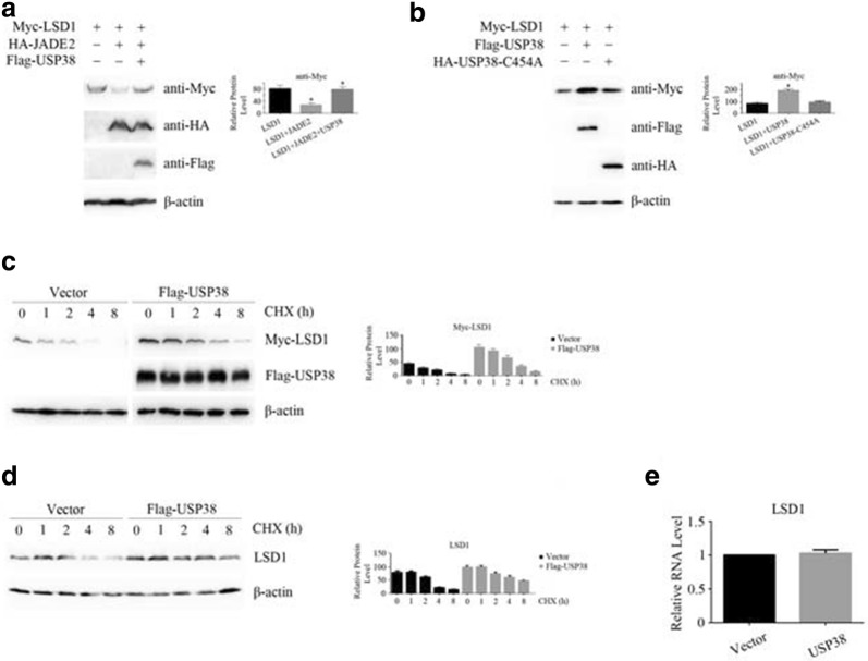
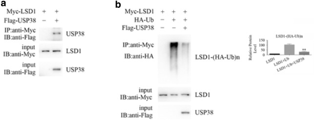
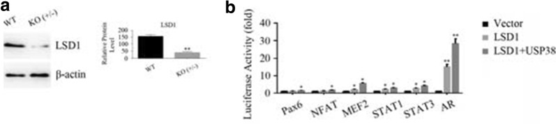
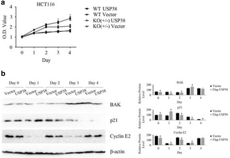
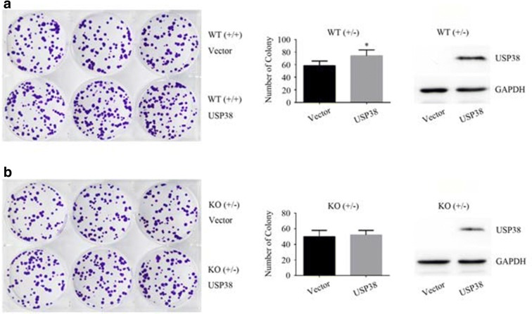
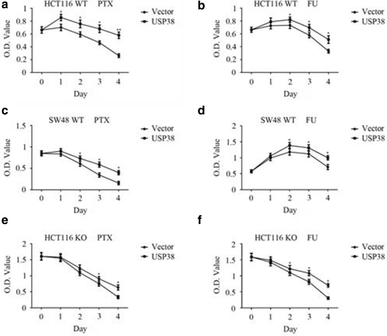
Results: The deubiquitinase USP38 stabilizes protein LSD1 in cells by binding LSD1 and cleaving its ubiquitin chain to prevent the degradation of LSD1 by the intracellular proteasome. USP38 enhances the ability of LSD1 to activate signaling pathways and hence promotes cellular abilities of proliferation and colony formation through interacting with LSD1. Furthermore, USP38 enhances the drug tolerance of human colon cancer cells.

Conclusions: USP38 is an LSD1-specific deubiquitinase that affects cellular physiology through interacting with LSD1.

Keywords: Deubiquitinase; LSD1; Proliferation; USP38.


Figures

Fig. 1
Fig. 1
The LSD1 protein is stabilized by the deubiquitinase USP38. a After tagged proteins of LSD1, JADE2 and USP38 proteins were expressed in HEK293T cells, the change in the LSD1 protein level was measured by western blotting. An asterisk (*) indicates p < 0.05, compared to LSD1-expressing or LSD1 and JADE2-expressing cells. b After LSD1 and USP38 or the USP38-C454A mutant were expressed in HEK293T cells, the change in the LSD1 protein level was measured by western blotting. An asterisk (*) indicates p < 0.05, compared to LSD1-expressing cells. c The Myc-tagged LSD1 gene was transfected into HEK293T cells together with the vector or Flag-tagged USP38 gene, and the cells were treated with cycloheximide (CHX) for 8 h. Western blotting was then performed to detect the changes in the protein level of Myc-LSD1. d The vector or Flag-tagged USP38 gene was transfected into HEK293T cells and the cells were treated with CHX for 8 h. Western blotting was then performed to detect the changes in the protein level of endogenously expressed LSD1. e Total RNAs were extracted from the vector- and Flag-USP38-transfected HEK293T cells and real-time quantitative PCR was performed to detect the changes in the LSD1 mRNA level. Three independent experiments were performed
Fig. 2
Fig. 2
USP38 binds LSD1 and removes its ubiquitin chain. a Coimmunoprecipitation was performed to detect the interaction between LSD1 and USP38. b Denaturing immunoprecipitation was applied to confirm that USP38 removed the ubiquitin chain from the LSD1 protein specifically. Double asterisks (**) indicate p < 0.01, compared to LSD1- and ubiquitin-expressing cells. Three independent experiments were performed
Fig. 3
Fig. 3
USP38 enhances the activation of signaling pathways activated by LSD1. a The protein levels of LSD1 in wild-type (WT, +/+) and single-allele knockout (KO, +/−) HCT116 cells. Double asterisks (**) indicate p < 0.01, compared to wild-type cells. b After the Myc-tagged LSD1 gene was transfected into HEK293T cells, luciferase assays were performed to detect the activation of signaling pathways. In addition, after Myc-tagged LSD1 and/or Flag-tagged USP38 genes were transfected into HEK293T cells, luciferase assays were performed to detect the activation of signaling pathways as well. An asterisk (*) indicates p < 0.05 and double asterisks (**) indicate p < 0.01, compared to vector-transfected cells. Three independent experiments were performed
Fig. 4
Fig. 4
USP38 promotes the proliferation of HCT116 cells. a After the vector or Flag-tagged USP38 gene was transfected into LSD1 wild-type (WT, +/+) or single-allele knockout (KO, +/−) HCT116 cells, cell proliferation assays were performed with CCK-8 kits. An asterisk (*) indicates p < 0.05, compared to vector-transfected HCT116 wild-type cells. b After the vector or Flag-tagged USP38 gene was transfected into LSD1 wild-type (WT, +/+) HCT116 cells, western blotting was performed to detect the expression of cell cycle and apoptosis related proteins. An asterisk (*) indicates p < 0.05, compared to vector-transfected cells. Three independent experiments were performed
Fig. 5
Fig. 5
Colony formation analysis. a LSD1 wild-type HCT116 cells (WT, +/+) were transfected with packaged lentiviruses of pHAGE-6tag or pHAGE-6tag-Flag-USP38, the stable USP38-expressing cell lines were screened with puromycin, and a colony formation assay was performed for 10 days. An asterisk (*) indicates p < 0.05, compared to vector-transfected cells. b LSD1 single-allele knockout (KO, +/−) HCT116 cells were transfected with packaged lentiviruses of pHAGE-6tag or pHAGE-6tag-Flag-USP38 and screened with puromycin, a colony formation assay was then performed for 13 days. Three independent experiments were performed
Fig. 6
Fig. 6
Drug tolerance assay. a HCT116 cells (LSD1 WT, +/+) stably expressing the USP38 protein and vector-transfected HCT116 cells (LSD1 WT, +/+) were treated with 40 ng/mL paclitaxel (PTX) for 4 days and CCK-8 was applied to measure cell proliferation. An asterisk (*) indicates p < 0.05 and double asterisks (**) indicate p < 0.01, compared to vector-transfected cells. b HCT116 cells (LSD1 WT, +/+) stably expressing the USP38 protein and vector-transfected HCT116 cells (LSD1 WT, +/+) were treated with 12.5 µg/mL 5-fluorouracil (5-FU) for 4 days and CCK-8 was applied to measure cell proliferation. An asterisk (*) indicates p < 0.05, compared to vector-transfected cells. c USP38 was expressed in SW48 cells (LSD1 WT, +/+), the cells were treated with PTX, and cell proliferation was analyzed. An asterisk (*) indicates p < 0.05, compared to vector-transfected cells. d USP38 was expressed in SW48 cells (LSD1 WT, +/+), the cells were treated with 5-FU, and cell proliferation was analyzed. An asterisk (*) indicates p < 0.05, compared to vector-transfected cells. e LSD1 single-allele knockout (KO, +/−) HCT116 cells were applied to be treated with PTX and cell proliferation was analyzed. An asterisk (*) indicates p < 0.05, compared to vector-transfected cells. f LSD1 single-allele knockout (KO, +/−) HCT116 cells were applied to be treated with 5-FU and cell proliferation was analyzed. An asterisk (*) indicates p < 0.05, compared to vector-transfected cells. Three independent experiments were performed

Similar articles

Cited by

References

    1. Coyne ES, Wing SS. The business of deubiquitination—location, location, location. F1000Res. 2016 doi: 10.12688/f1000research.7220.1. - DOI - PMC - PubMed
    1. Eletr ZM, Wilkinson KD. Regulation of proteolysis by human deubiquitinating enzymes. Biochim Biophys Acta. 2014;1843(1):114–128. doi: 10.1016/j.bbamcr.2013.06.027. - DOI - PMC - PubMed
    1. Reyes-Turcu FE, Ventii KH, Wilkinson KD. Regulation and cellular roles of ubiquitin-specific deubiquitinating enzymes. Annu Rev Biochem. 2009;78:363–397. doi: 10.1146/annurev.biochem.78.082307.091526. - DOI - PMC - PubMed
    1. Suresh B, Lee J, Kim KS, Ramakrishna S. The importance of ubiquitination and deubiquitination in cellular reprogramming. Stem Cells Int. 2016 doi: 10.1155/2016/6705927. - DOI - PMC - PubMed
    1. Choi J, Jang H, Kim H, Kim ST, Cho EJ, Youn HD. Histone demethylase LSD1 is required to induce skeletal muscle differentiation by regulating myogenic factors. Biochem Biophys Res Commun. 2010;401(3):327–332. doi: 10.1016/j.bbrc.2010.09.014. - DOI - PubMed

MeSH terms