Review
. 2019 Feb;29(2):135-149.
doi: 10.1016/j.tcb.2018.10.006. Epub 2018 Nov 26.

Homologous Recombination and the Formation of Complex Genomic Rearrangements

Affiliations
Review

Homologous Recombination and the Formation of Complex Genomic Rearrangements

Aurèle Piazza et al. Trends Cell Biol. 2019 Feb.

Abstract

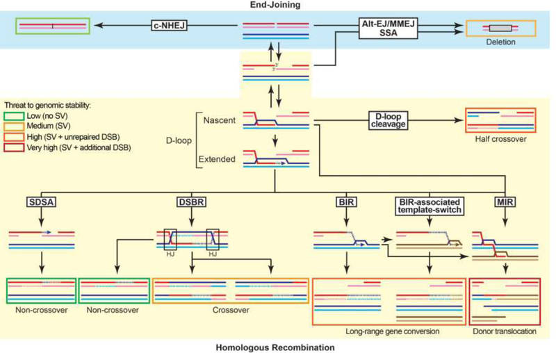
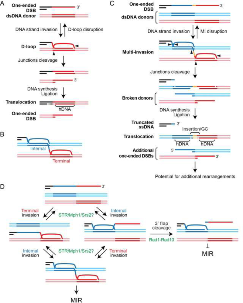
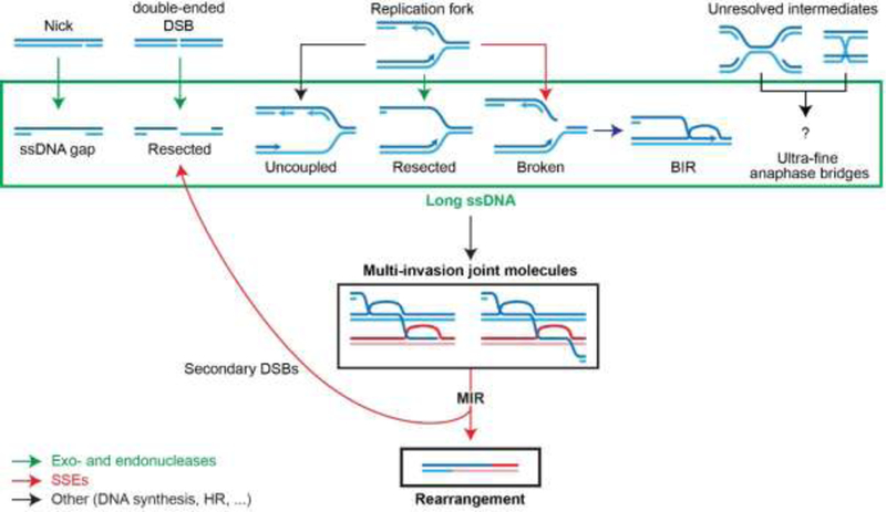
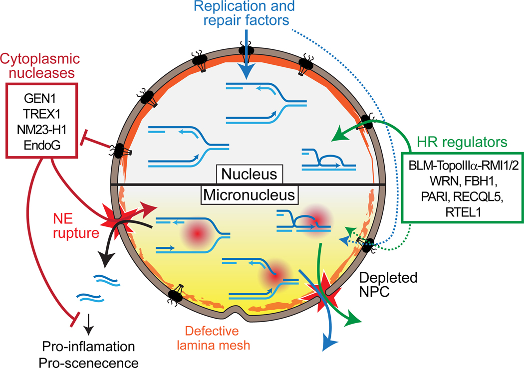
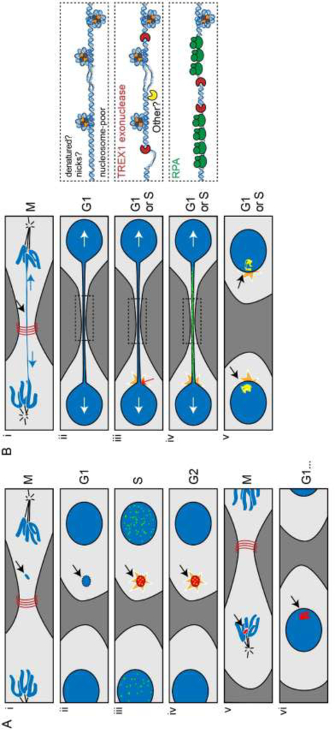
The maintenance of genome integrity involves multiple independent DNA damage avoidance and repair mechanisms. However, the origin and pathways of the focal chromosomal reshuffling phenomena collectively referred to as chromothripsis remain mechanistically obscure. We discuss here the role, mechanisms, and regulation of homologous recombination (HR) in the formation of simple and complex chromosomal rearrangements. We emphasize features of the recently characterized multi-invasion (MI)-induced rearrangement (MIR) pathway which uniquely amplifies the initial DNA damage. HR intermediates and cellular contexts that endanger genomic stability are discussed as well as the emerging roles of various classes of nucleases in the formation of genome rearrangements. Long-read sequencing and improved mapping of repeats should enable better appreciation of the significance of recombination in generating genomic rearrangements.

Keywords: chromothripsis; copynumber variation; multi-invasion; non-allelic homologous recombination; structural variant; structure-selective endonuclease.


Figures

Figure 1:
Figure 1:. Overview of the DSB repair pathways and their associated risk for genomic stability.
The repair products are boxed. The box color (from green to red) indicates the threat to genomic stability posed by the product of each pathway and sub-pathway. DNA synthesis is indicated by an arrow and newly-synthesized DNA by a dotted line. The long ssDNA associated with BIR has the potential to undergo MIR. More detailed mechanisms for D-loop cleavage and MIR are provided in Figures 2A and 2C, respectively. The reversibility of DSB resection provided by the Shieldin complex with fill-in by the DNA polymerase α-primase complex provides an unanticipated degree of flexibility in the choice of DSB repair between HR and EJ mechanisms [130]. Hence, EJ pathways are available not only on unresected or minimally resected DSB, they can also be engaged after extensive resection and subsequent fill-in.
Figure 2:
Figure 2:. Single and multi-invasion joint molecules are at-risk HR intermediates for SSE- mediated genomic instability.
(A) Model for half-crossover formation from cleavage of a single D-loop. (B) Multi-invasions (MI) joint molecules are formed when a Rad51-ssDNA filament invades two independent donors along its length. It features an internal and a terminal D-loop. (C) Model for multi-invasion-induced rearrangement (MIR). (D) Several activities inhibit MIR in a reversible (Sgs1-Top2-Rmi1 (STR), Mph1, Srs2) or irreversible (Rad1-Rad10) fashion. The specificity of STR, Mph1 and Srs2 as well as the precise nature of their substrates has not been established.
Figure 3:
Figure 3:. Nuclease involvement in MIR.
Exo- and endonucleases involved in resection (green) generate long recombinogenic substrates, while SSEs (red) process MI joint molecules into rearrangements and additional resected secondary single-ended DSBs (indicated by backwards arrow to resected DSBs).
Figure 4:
Figure 4:. S-phase-specific DNA damage from defective nucleoplasm isolation and DNA metabolism in micronuclei.
The nuclear envelope (NE) of micronuclei is depleted for nuclear pore complex and features abnormal lamina deposition. These dysregulations lead to abnormal nuclear protein import and frequent NE rupture, respectively. NE rupture leads to the penetration of cytoplasmic components to the micronucleus and the leakage of soluble nuclear components and DNA.
Figure 5:
Figure 5:. Cellular models for chromothripsis.
(A) Chromothripsis by isolation isolation and breakage of chromosomes in micronuclei [92, 93]. (i-ii) Mis-segregation and physical separation of a large chromatin fragment leads to micronucleus formation. (ii-iii) Micronuclei are defective for the nuclear import and barrier function (Fig. 4). (iii) Upon S-phase entry, NE rupture leads to the formation of massive DNA damage, which features co-localizing DSBs (green) and γH2AX foci [95] (iv-v) The micronuclear DNA reaches mitosis under-replicated and fragmented. (v-vi) Micronuclear DNA fragments can be re-incorporated into the main nucleus at subsequent mitosis where it may undergo final repair. (B) Chromothripsis originating from attempted segregation of dicentric chromosomes [94]. (i) Spindle-exerted tensions can stretch but not break the central portion of a dicentric chromosome. (ii) NE forms around the daughter nuclei and the intervening chromatin bridge, depleted for lamin and nuclear pore complex. Right inset: the bridge is partially depleted for nucleosomes. (iii) Tensions exerted on the bridge cause NE rupture at the base of the bridge in G1/S phase. NE rupture leads to leaking of nuclear components and penetration of cytoplasmic proteins such as the TREX1 exonuclease (right inset). (iv) TREX1 exploits nicks or ssDNA gaps present in the bridge or generated by other cytoplasmic endonuclease to resect the chromatin bridge (right inset). This leads to accumulation of RPA foci (green) and (v) culminates in the resolution of the bridge. In addition to RPA, the resected DNA causes the formation of 53BP1, γH2AX and Mre11 foci (yellow) in the daughter nuclei.

References

    1. Stephens PJ et al. (2011) Massive genomic rearrangement acquired in a single catastrophic event during cancer development. Cell 144, 27–40. - PMC - PubMed
    1. Cortes-Ciriano I et al. (2018) Comprehensive analysis of chromothripsis in 2,658 human cancers using whole-genome sequencing. bioRxiv - PMC - PubMed
    1. Kloosterman WP and Cuppen E (2013) Chromothripsis in congenital disorders and cancer: similarities and differences. Curr Opin Cell Biol 25, 341–8. - PubMed
    1. Liu P et al. (2011) Chromosome catastrophes involve replication mechanisms generating complex genomic rearrangements. Cell 146, 889–903. - PMC - PubMed
    1. Korbel JO and Campbell PJ (2013) Criteria for inference of chromothripsis in cancer genomes. Cell 152, 1226–36. - PubMed

Publication types