. 2018 Dec:38:79-88.
doi: 10.1016/j.ebiom.2018.11.030. Epub 2018 Nov 27.

Ectopic high endothelial venules in pancreatic ductal adenocarcinoma: A unique site for targeted delivery

Affiliations

Ectopic high endothelial venules in pancreatic ductal adenocarcinoma: A unique site for targeted delivery

Baharak Bahmani et al. EBioMedicine. 2018 Dec.

Abstract

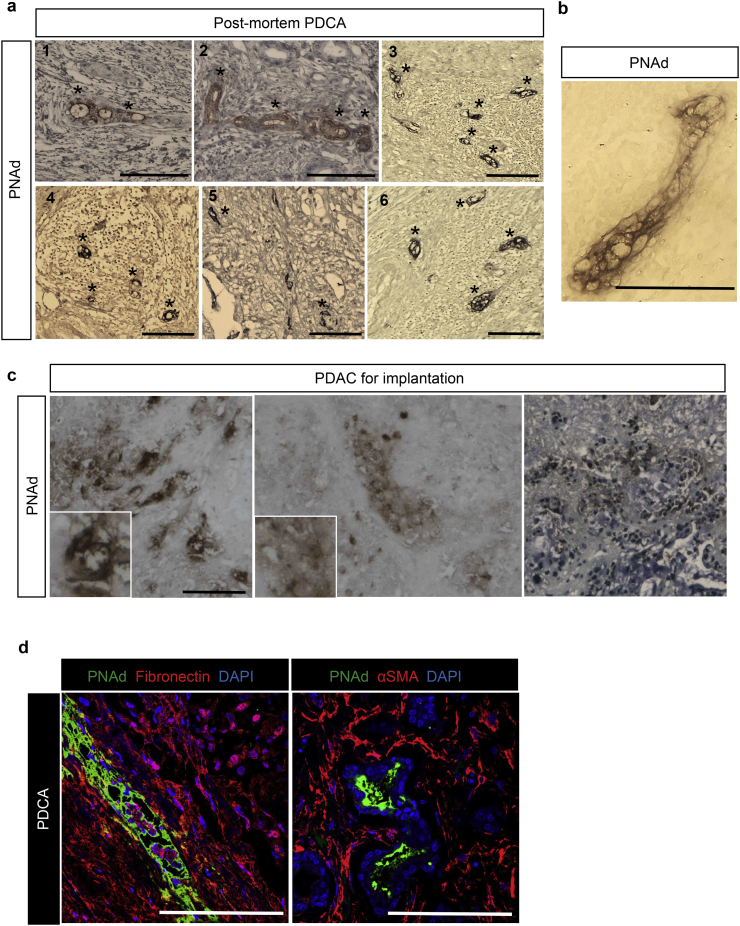
Background: Nanomedicine offers an excellent opportunity to tackle treatment-refractory malignancies by enhancing the delivery of therapeutics to the tumor site. High endothelial venules (HEVs) are found primarily in lymph nodes or formed de novo in peripheral tissues during inflammatory responses. They express peripheral node addressin (PNAd), which is recognized by the monoclonal antibody MECA79.

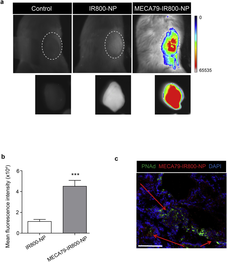
Methods: Here, we demonstrated that HEVs form de novo in human pancreatic ductal adenocarcinoma (PDAC). We engineered MECA79 coated nanoparticles (MECA79-NPs) that recognize these ectopic HEVs in PDAC.

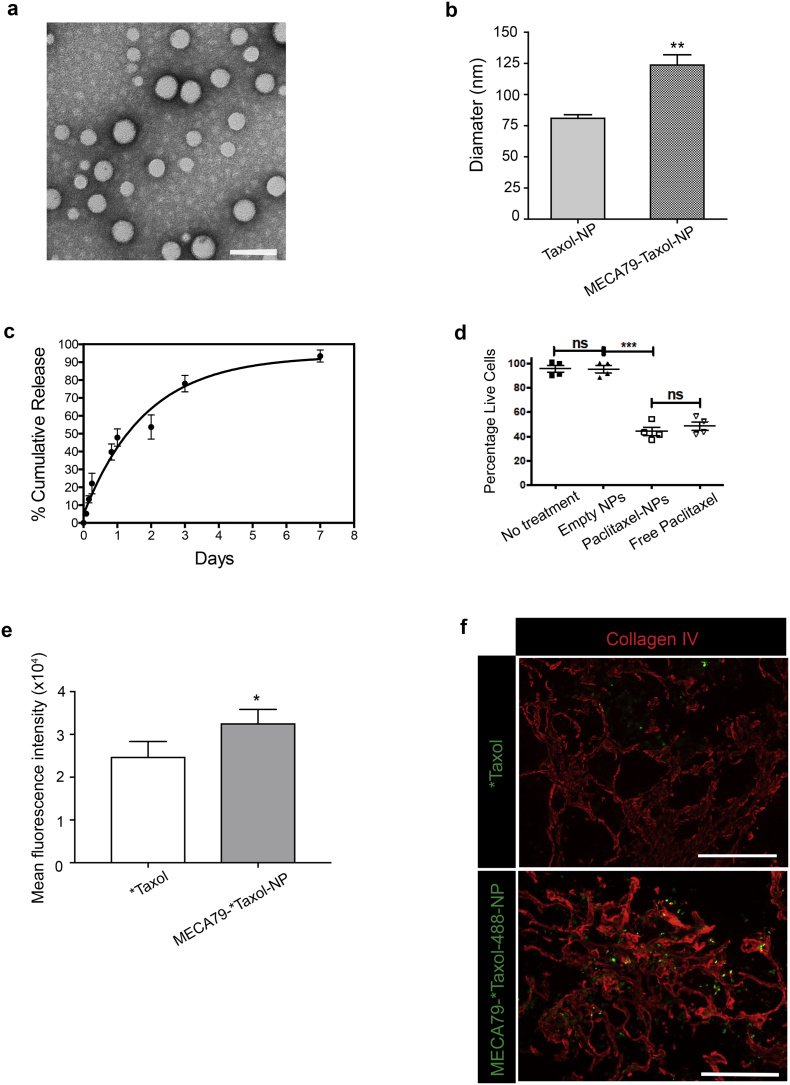
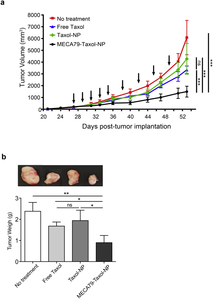
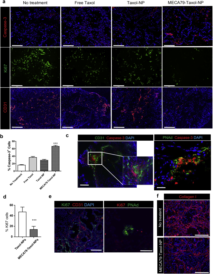
Findings: The trafficking of MECA79-NPs following intravenous delivery to human PDAC implanted in a humanized mouse model was more robust than non-conjugated NPs. Treatment with MECA79-Taxol-NPs augmented the delivery of Paclitaxel (Taxol) to the tumor site and significantly reduced the tumor size. This effect was associated with a higher apoptosis rate of PDAC cells and reduced vascularization within the tumor.

Interpretation: Targeting the HEVs of PDAC using MECA79-NPs could lay the ground for the localized delivery of a wide variety of drugs including chemotherapeutic agents. FUND: National Institutes of Health (NIH) grants: T32-EB016652 (B·B.), NIH Cancer Core Grant CA034196 (L.D.S.), National Institute of Allergy and Infectious Diseases grants R01-AI126596 and R01-HL141815 (R.A.).

Keywords: High endothelial venules; MECA79 coated nanoparticles; Pancreatic ductal adenocarcinoma; Peripheral node addressin; Taxol.


Figures

Fig. 1
Fig. 1
Immunohistochemistry and immunofluorescence analysis of human PDAC. (a) Representative images from immunohistochemistry analysis of human PDAC samples from post-mortem showing ectopic PNAd+ venules (marked by * in the images, n = 6, scale bar = 100 μm). (b) A representative high-magnification IHC image of a PNAD+ vessel in the PDAC (Scale bar = 75 μm). (c) Immunohistochemistry staining of the human PDAC prior to implantation into the NSG mice confirming PNAd expression in the tissue. (d) Representative images of PNAD+ vessels (green) surrounded by Fibronectin (red) and αSMA (red) (Scale bar = 100 μm).
Fig. 2
Fig. 2
Targeted delivery of NP to the PNAd-expressing HEV in PDAC. (a) Whole-body NIR fluorescent imaging showing higher trafficking of MECA79-IR800-NPs to PDAC, as compared to non-conjugated IR800-NPs. The dashed area in each image represents the PDAC tumor in vivo and ex vivo post-harvest, respectively. (b) Corresponding MFI of the tumor showing a significant increase in trafficking of MECA79-IR800-NPs, as compared to the non-conjugated IR800-NPs. The data are presented as the mean and SEM (n = 3). (***p < 0.001, student's t-test). (c) Representative immunofluorescence micrograph of the tumor 24 h post-injection of MECA79-IR800-NPs (red) demonstrates their presence around HEV (green). The red arrows point to the areas of MECA79-IR800-NP accumulation in the tumor (Scale bar = 100 μm).
Fig. 3
Fig. 3
Formulation and characterization of MECA79-Taxol-NPs. (a) Representative TEM images of Taxol-NP (Scale bar = 100 nm) (b) Hydrodynamic diameter of non-conjugated and MECA79-conjugated Taxol-NPs. The data are presented as the mean and SEM (n = 6). (c) Release kinetics of Taxol from NPs over one-week. The data are presented as the mean and SEM (n = 3). (d) Taxol-NPs induced similar levels of apoptosis in pancreatic cancer cells in vitro in comparison to free Taxol. (e) The MFI of tumors showed significantly higher accumulation of *Taxol in MECA79-*Taxol-NP group, as compared to *Taxol group (*p < 0.05, student's t-test). The data are presented as the mean and SEM (n = 3). (f) Immunofluorescence staining of PDAC tissue showed that MECA79-*Taxol-NPs (green) were present within the ECM (Collagen IV, in red) of tumor at a higher number, as compared to the control group (Scale bar = 100 μm).
Fig. 4
Fig. 4
Therapeutic efficacy of MECA79-Taxol-NPs. (a) Improved efficacy in delivering Taxol to PDAC was observed in mice treated with MECA79-Taxol-NPs, as compared to no treatment, free Taxol, and Taxol-NPs (Taxol dose =0.5 mg/kg, ***p < 0.001, ANOVA, n = 5 mice/group). Arrows indicate the days of treatment. The data are presented as the mean and SEM. (b) The final weight of the tumor in mice treated with MECA79-Taxol-NPs was significantly lower, as compared to the no treatment, free Taxol, and Taxol-NP treatment groups. (*p < 0.05, **p < 0.01, ANOVA, n = 5 mice/group). The data are presented as the mean and SEM.
Fig. 5
Fig. 5
Histological analysis of PDAC following treatment with MECA79-Taxol-NPs. (a) Immunofluorescence staining of the tumor at the end of the study demonstrated that treatment with MECA79-Taxol-NPs resulted in higher apoptosis (caspase-3+) of cancer cells, lower cellular proliferation (Ki67+), and decreased vascularization (CD31+) (Scale bar = 200 μm). (b) Quantification of the caspase-3+ cells using ImageJ software (***p < 0.001, ANOVA, n = 3 mice/group). The data are presented as the mean and SEM. (c) Representative immunofluorescence images of the PDAC tumor for markers of vasculature (CD31 and PNAd in green) and apoptosis (caspase-3 in red) following treatment with MECA79-Taxol-NPs (Scale bar = 200 μm). (d) Quantification of the Ki67+ cells using ImageJ software (***p < 0.001, student's t-test, n = 3 mice/group). The data are presented as the mean and SEM. (e) Representative immunofluorescence images of the PDAC tumor for markers of vasculature (CD31 and PNAd) and proliferation (Ki67) in the no-treatment group (Scale bar = 200 μm for CD31, 100 μm for PNAd). (f) Immunofluorescence staining for collagen I (red) of the PDAC tumor comparing the groups that received no treatment and MECA79-Taxol-NP (Scale bar = 100 μm).

Comment in

References

    1. Siegel R.L., Miller K.D., Jemal A. Cancer statistics, 2016. CA Cancer J Clin. 2016;66(1):7–30. - PubMed
    1. Kleeff J., Korc M., Apte M., La Vecchia C., Johnson C.D., Biankin A.V. Pancreatic cancer. Nat Rev Dis Prim. 2016;2:16022. - PubMed
    1. Ying H., Dey P., Yao W., Kimmelman A.C., Draetta G.F., Maitra A. Genetics and biology of pancreatic ductal adenocarcinoma. Genes Dev. 2016;30(4):355–385. - PMC - PubMed
    1. Ryan D.P., Hong T.S., Bardeesy N. Pancreatic Adenocarcinoma. N Engl J Med. 2014;371(11):1039–1049. - PubMed
    1. Chiaravalli M., Reni M., O'Reilly E.M. Pancreatic ductal adenocarcinoma: State-of-the-art 2017 and new therapeutic strategies. Cancer Treat Rev. 2017;60:32–43. - PubMed

MeSH terms