. 2018 Dec 18;115(51):13099-13104.
doi: 10.1073/pnas.1808137115. Epub 2018 Nov 29.

Removal of clock gene Bmal1 from the retina affects retinal development and accelerates cone photoreceptor degeneration during aging

Affiliations

Removal of clock gene Bmal1 from the retina affects retinal development and accelerates cone photoreceptor degeneration during aging

Kenkichi Baba et al. Proc Natl Acad Sci U S A. .

Abstract

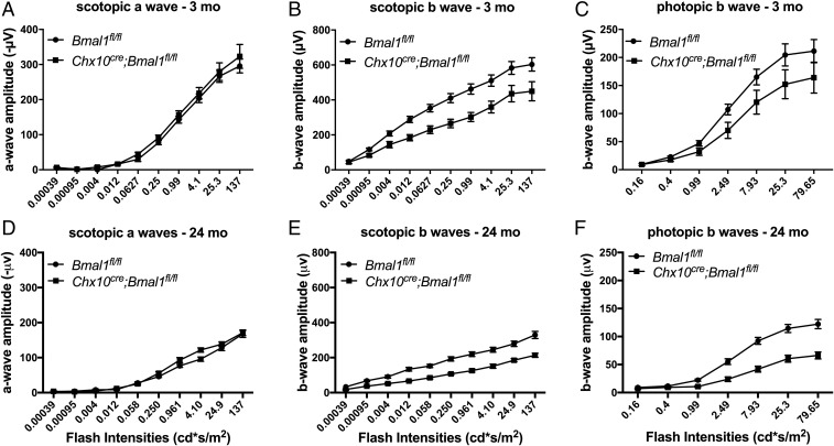
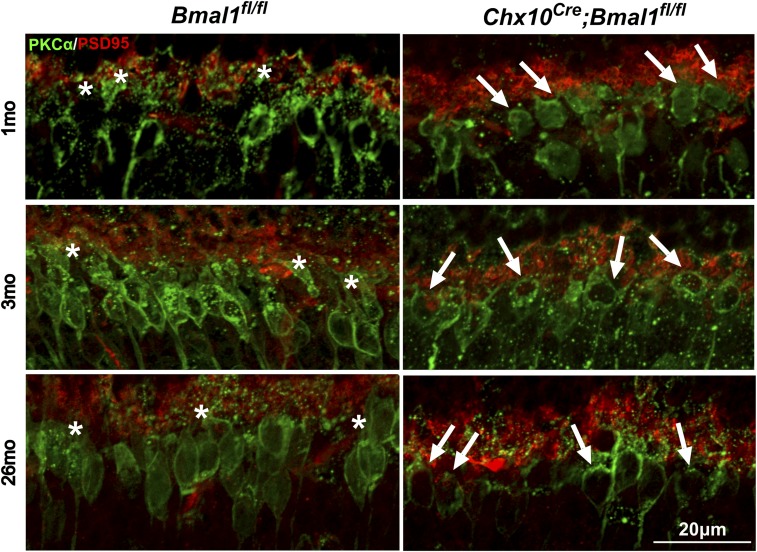
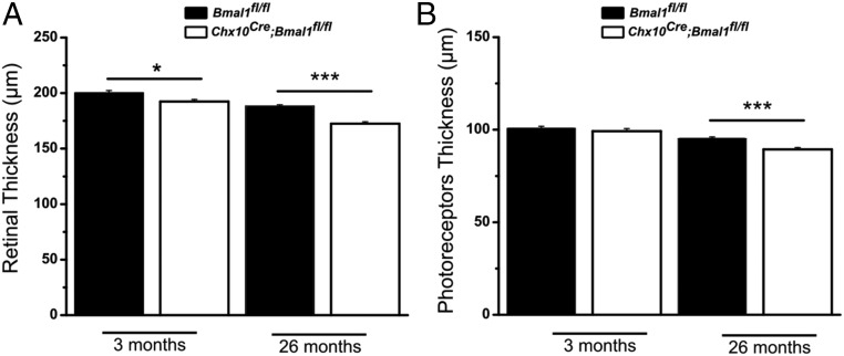
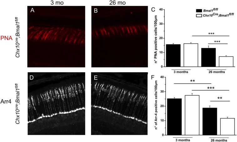
The mammalian retina contains an autonomous circadian clock system that controls many physiological functions within this tissue. Previous studies on young mice have reported that removal of the key circadian clock gene Bmal1 from the retina affects the circadian regulation of visual function, but does not affect photoreceptor viability. Because dysfunction in the circadian system is known to affect cell viability during aging in other systems, we compared the effect of Bmal1 removal from the retina on visual function, inner retinal structure, and photoreceptor viability in young (1 to 3 months) and aged (24 to 26 months) mice. We found that removal of Bmal1 from the retina significantly affects visual information processing in both rod and cone pathways, reduces the thickness of inner retinal nuclear and plexiform layers, accelerates the decline of visual functions during aging, and reduces the viability of cone photoreceptors. Our results thus suggest that circadian clock dysfunction, caused by genetic or other means, may contribute to the decline of visual function during development and aging.

Keywords: aging; circadian rhythm; cone photoreceptor; development; retina.


Conflict of interest statement

The authors declare no conflict of interest.

Figures

Fig. 1.
Fig. 1.
Scotopic and photopic ERGs in Chx10Cre;Bmal1fl/fl and Bmal1fl/fl mice. Scotopic and photopic ERGs were obtained from young (AC) and old (DF) mice. The scotopic and photopic b waves, but not a waves, were affected in Chx10Cre;Bmal1fl/fl mice compared with littermate Bmal1fl/fl controls (P < 0.05; n = 5 to 8). The amplitudes of both scotopic and photopic ERGs decline with age (P < 0.01), and there was a trend for this decline to be more pronounced in Chx10Cre;Bmal1fl/fl mice compared with Bmal1fl/fl mice.
Fig. 2.
Fig. 2.
The dendritic processes of rod bipolar cells are affected in Chx10Cre;Bmal1fl/fl mice. Synaptic connections between photoreceptor cells (anti-PSD95, red) and bipolar cells (anti-PKCα, green) in Chx10Cre;Bmal1fl/fl and Bmal1fl/fl mice at the ages of 1, 3, and 26 mo. White asterisks show the presence of dendritic arborization in Bmal1fl/fl mice; white arrows show the stunted dendritic arborizations in Chx10Cre;Bmal1fl/fl mice at all of the ages tested.
Fig. 3.
Fig. 3.
Quantification of SD-OCT. (A) A small but significant difference in the total thickness of the retina was detected between Bmal1fl/fl and Chx10Cre;Bmal1fl/fl genotypes at both ages investigated (*P < 0.05 at 3 mo and ***P < 0.001 at 24 mo). (B) No difference in the thickness of PRL was observed between young Bmal1fl/fl and Chx10Cre;Bmal1fl/fl mice, whereas a significant reduction in the thickness of the PRL was detected in older Chx10Cre;Bmal1fl/fl mice with respect to the Bmal1fl/fl mice (***P < 0.001 at 24 mo).
Fig. 4.
Fig. 4.
Morphological evaluation of cone photoreceptors in young and old Bmal1fl/fl and Chx10Cre;Bmal1fl/fl mice. Peanut agglutinin (PNA) labeling in the central retina of young (A) and old (B) Chx10Cre;Bmal1fl/fl mice. (C) Quantification of PNA-labeled cones of Bmal1fl/fl and Chx10Cre;Bmal1fl/fl mice retinas at 3 and 26 mo of age. A significant change in the number of PNA-positive cells is observed between Bmal1fl/fl and Chx10Cre;Bmal1fl/fl mice at 26 mo of age (***P < 0.001). Cone arrestin (Arr4) staining in young (D) and old (E) Chx10Cre;Bmal1fl/fl mice. (F) Quantification of cone arrestin staining in Bmal1fl/fl and Chx10Cre;Bmal1fl/fl mice retinas at 3 and 26 mo of age. A significant change in the number of arrestin-positive cells is observed between Bmal1fl/fl and Chx10Cre;Bmal1fl/fl mice at 26 mo of age (**P < 0.01). A significant reduction in the number of cones is also observed in both genotypes during aging (Bmal1fl/fl, **P < 0.01; Chx10Cre;Bmal1fl/fl, ***P < 0.001). Each bar represents the mean ± SEM (n = 3 to 4).

References

    1. Takahashi JS. Transcriptional architecture of the mammalian circadian clock. Nat Rev Genet. 2017;18:164–179. - PMC - PubMed
    1. Tosini G, Menaker M. Circadian rhythms in cultured mammalian retina. Science. 1996;272:419–421. - PubMed
    1. Ruan G-X, Zhang DQ, Zhou T, Yamazaki S, McMahon DG. Circadian organization of the mammalian retina. Proc Natl Acad Sci USA. 2006;103:9703–9708. - PMC - PubMed
    1. Tosini G, Davidson AJ, Fukuhara C, Kasamatsu M, Castanon-Cervantes O. Localization of a circadian clock in mammalian photoreceptors. FASEB J. 2007;21:3866–3871. - PMC - PubMed
    1. Ruan G-X, Allen GC, Yamazaki S, McMahon DG. An autonomous circadian clock in the inner mouse retina regulated by dopamine and GABA. PLoS Biol. 2008;6:e249. - PMC - PubMed

Publication types

Substances

LinkOut - more resources