. 2018 Oct 24:12:2167-2176.
doi: 10.2147/OPTH.S174573. eCollection 2018.

Clinical spectrum of severe chronic central serous chorioretinopathy and outcome of photodynamic therapy

Affiliations

Clinical spectrum of severe chronic central serous chorioretinopathy and outcome of photodynamic therapy

Danial Mohabati et al. Clin Ophthalmol. .

Abstract

Purpose: To describe a spectrum of severe chronic central serous chorioretinopathy (cCSC) cases and their response to photodynamic therapy (PDT).

Patients and methods: A total of 66 patients (81 eyes) with active severe cCSC were studied, and their response to PDT was compared with a control group consisting of 35 active cCSCs (37 eyes) that did not display characteristics of severity. Best-corrected visual acuity (BCVA) and complete resolution of subretinal fluid (SRF) were considered as main outcome measures.

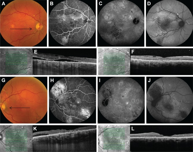
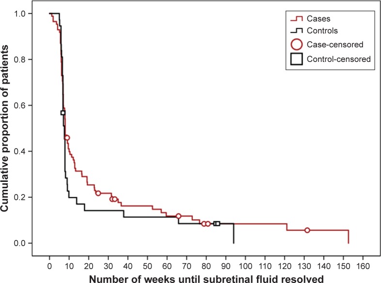
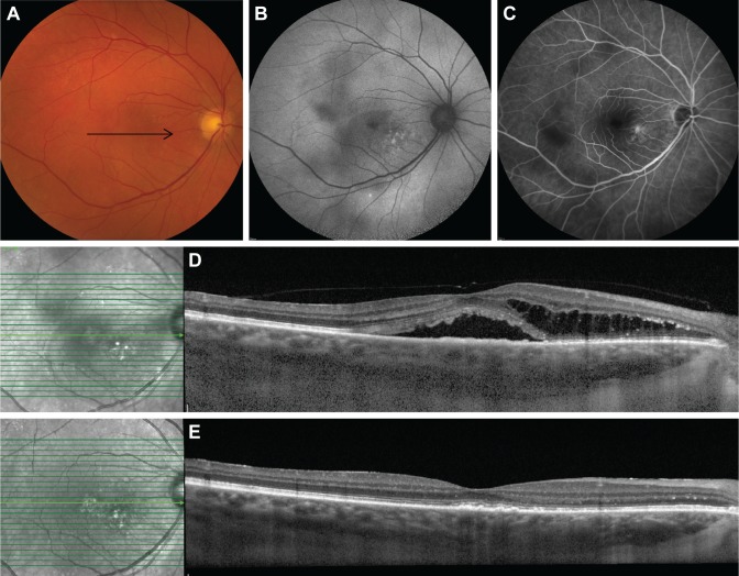
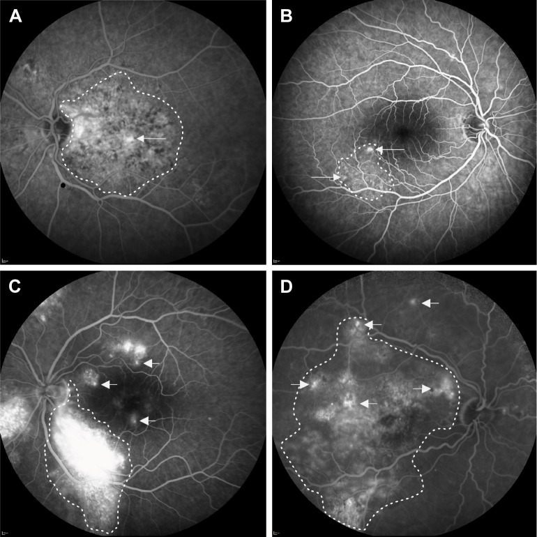
Results: In severe cCSC cases, we found cumulative areas of diffuse atrophic retinal pigment epithelium alterations in 48 eyes (59%), multiple "hot spots" of leakage in 36 eyes (44%), posterior cystoid retinal degeneration in 25 eyes (31%), and 13 eyes (16%) had a diffuse leakage on fluorescein angiography. After PDT treatment, BCVA increased in both groups, from 66 to 72 Early Treatment of Diabetic Retinopathy Study (ETDRS) letters in the case group (P<0.001), and from 78 to 82 ETDRS letters in the control group (P<0.001). SRF had resolved completely in 87% of severe cCSC cases and 95% of controls at final follow-up visit.

Conclusion: A spectrum of severe cCSC exists, and PDT seems to be an effective treatment in both severe cCSC and nonsevere cCSC in terms of resolution of SRF. Final BCVA shows a significant improvement in both groups after PDT treatment.

Keywords: chronic central serous chorioretinopathy; photodynamic therapy; posterior cystoid retinal degeneration; severe phenotype; therapeutic outcome.


Conflict of interest statement

Disclosure The authors report no conflicts of interest in this work.

Figures

Figure 1
Figure 1
Multimodal imaging of a 71-year-old male patient with severe bilateral chronic central serous chorioretinopathy (AF: right eye, GL: left eye). Notes: On color fundus photography, atrophic RPE alterations were seen in the inferotemporal quadrant of the left eye (G). Multifocal “hot spots” of leakage (H) and extensive areas of atrophy were seen on fluorescein angiography (B, H). Fundus autofluorescence showed large areas of hypo- and hyper-autofluorescent abnormalities corresponding to the RPE changes, extending to outside the macula (D, J). Indocyanine green angiography images (C, I) showed multifocal areas of diffuse choroidal abnormalities and leakage. OCT revealed in both eyes epiretinal membrane and subretinal fluid centrally in the macula (E, K). Posterior cystoid retinal degeneration was seen in the outer nuclear layer of the nasal macula of the left eye (K). Ten weeks after half-dose photodynamic therapy, which was only performed in the left eye, both subretinal fluid and posterior cystoid retinal degeneration had disappeared (L). The black arrows on the color fundus photography images correspond to the scanning plane on the OCT scans (E, F, K, L). Abbreviations: OCT, optical coherence tomography; RPE, retinal pigment epithelial.
Figure 2
Figure 2
Kaplan–Meier curve showing the cumulative fraction of patients treated for chronic central serous chorioretinopathy (cCSC). Notes: Endpoint: “Complete resolution of subretinal fluid (SRF) after photodynamic therapy”; the median duration to SRF resolution in cCSC patients with a severe phenotype of the disease (cases) was 8 weeks (95% CI: 7–9). In cCSC patients who did not meet the criteria of severity (controls), the median duration was 7 weeks (95% CI: 7–8; log-rank test; P=0.281).
Figure 3
Figure 3
Clinical features on multimodal imaging of the right eye of a 63-year-old female patient with severe chronic central serous chorioretinopathy (AC). Black arrow on color fundus photography image (A) shows the scanning plane, which is depicted on the SD-OCT scans (D, E). FAF shows multiple speckled hyperautofluorescent changes in the macula together with an irregular surface of hypoautofluorescence, expanding from the fovea to the superior and inferior vascular arcades (B). Fluorescein angiography imaging (C) revealed a limited area of fluorescein leakage with a clear central focus. The areas of hypofluorescence were located more temporal from the fovea and were smaller than on FAF. An SD-OCT scan (D) at first presentation and prior to treatment revealed a subretinal serous fluid accumulation together with a posterior cystoid retinal degeneration in the outer nuclear layer of the retina. At ~2 months after half-dose photodynamic therapy, both subretinal and intraretinal fluid on OCT had resolved completely (E). Abbreviations: FAF, fundus autofluorescence imaging; SD-OCT, spectral-domain optical coherence tomography.
Figure 4
Figure 4
Four categories of eyes with severe chronic central serous chorioretinopathy with active fluorescein leakage on fluorescein angiography. Notes: This figure illustrates different fluorescein leakage locations (arrows) in relation to the largest area of DARA (dotted line). (A) Category 1 concerns a leakage point inside the largest area of RPE alterations. (B) Category 2 concerns 1 or more leakage points located on the edges of the largest region of RPE alterations. (C) In category 3, leakage points causing macular subretinal fluid are outside the largest zone of RPE alterations. (D) Category 4 concerns cases with multifocal leakage points in several of the aforementioned locations in relation to the area of RPE alterations. Abbreviations: DARA, diffuse atrophic RPE alterations; RPE, retinal pigment epithelium.

References

    1. Gass JD. Pathogenesis of disciform detachment of the neuroepithelium. Am J Ophthalmol. 1967;63(3):1–139. - PubMed
    1. Nicholson B, Noble J, Forooghian F, Meyerle C. Central serous chorioretinopathy: update on pathophysiology and treatment. Surv Ophthalmol. 2013;58(2):103–126. - PMC - PubMed
    1. Liew G, Quin G, Gillies M, Fraser-Bell S. Central serous chorioretinopathy: a review of epidemiology and pathophysiology. Clin Exp Ophthalmol. 2013;41(2):201–214. - PubMed
    1. Fujimoto H, Gomi F, Wakabayashi T, et al. Morphologic changes in acute central serous chorioretinopathy evaluated by fourier-domain optical coherence tomography. Ophthalmology. 2008;115(9):1494–1500. 1500.e1–2. - PubMed
    1. Breukink MB, Dingemans AJ, den Hollander AI, et al. Chronic central serous chorioretinopathy: long-term follow-up and vision-related quality of life. Clin Ophthalmol. 2017;11:39–46. - PMC - PubMed