. 2018 Nov 28;4(11):eaat3834.
doi: 10.1126/sciadv.aat3834. eCollection 2018 Nov.

Accumulation of JAK activation loop phosphorylation is linked to type I JAK inhibitor withdrawal syndrome in myelofibrosis

Affiliations

Accumulation of JAK activation loop phosphorylation is linked to type I JAK inhibitor withdrawal syndrome in myelofibrosis

Denis Tvorogov et al. Sci Adv. .

Abstract

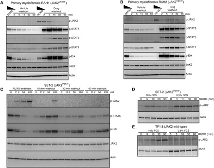
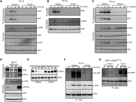
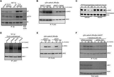
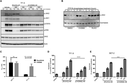
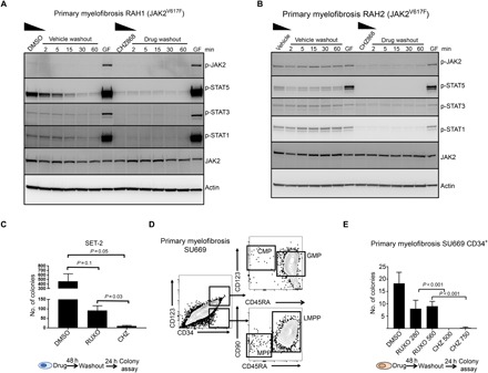
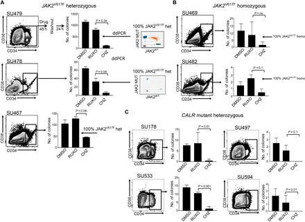
Treatment of patients with myelofibrosis with the type I JAK (Janus kinase) inhibitor ruxolitinib paradoxically induces JAK2 activation loop phosphorylation and is associated with a life-threatening cytokine-rebound syndrome if rapidly withdrawn. We developed a time-dependent assay to mimic ruxolitinib withdrawal in primary JAK2V617F and CALR mutant myelofibrosis patient samples and observed notable activation of spontaneous STAT signaling in JAK2V617F samples after drug washout. Accumulation of ruxolitinib-induced JAK2 phosphorylation was dose dependent and correlated with rebound signaling and the presence of a JAK2V617F mutation. Ruxolitinib prevented dephosphorylation of a cryptic site involving Tyr1007/1008 in JAK2 blocking ubiquitination and degradation. In contrast, a type II JAK inhibitor, CHZ868, did not induce JAK2 phosphorylation, was not associated with withdrawal signaling, and was superior in the eradication of flow-purified JAK2V617F mutant CD34+ progenitors after drug washout. Type I inhibitor-induced loop phosphorylation may act as a pathogenic signaling node released upon drug withdrawal, especially in JAK2V617F patients.


Figures

Fig. 1
Fig. 1. Ruxolitinib washout triggers intracellular signaling in patients with myelofibrosis.
(A and B) Mononuclear cells from patients with JAK2V617F myelofibrosis RAH1 and RAH2 were cultured for 12 hours in media containing 10% FCS and erythropoietin (EPO) and interleukin-3 (IL-3) (1 ng/ml each) with 280 nM ruxolitinib or dimethyl sulfoxide (DMSO) as vehicle control. Cells were washed in cold media and transferred into prewarmed and CO2-equilibrated media without additives for the indicated periods of time, and cell lysates were prepared and immunoblotted for the indicated proteins. (C) JAK2V617F positive SET-2 cells were cultured with ruxolitinib (0, 11.2, 56, or 280 nM) for 12 hours, followed by washout of drug. Whole-cell lysates were prepared at 0, 15, 30, and 60 min and analyzed by immunoblotting with p-JAK2, p-STAT5, and p-ERK antibodies. SET-2 (D) or TF1.8 (E) cells were incubated for 6 hours in media containing either 10 or 0.5% FCS before the addition of 280 nM ruxolitinib for 0, 5, 10, 30, and 90 min. Whole-cell lysates were prepared and immunoblotted with p-JAK2 and total JAK2 antibodies.
Fig. 2
Fig. 2. Type I JAK2 inhibitor protects JAK2 from degradation and down-regulation.
(A and B) TF1.8 cells were starved overnight in the presence of 0.5% FCS, preincubated with either vehicle or 280 nM ruxolitinib for 10 min, and stimulated with IL-3 (50 ng/ml) for different times. Cells were lysed and subjected to immunoprecipitation with (A) anti-phosphotyrosine (4G10) or (B) anti-phosphorylated JAK2 (Y1007/1008) antibodies, followed by immunoblotting with JAK2 and JAK1 antibodies. As a control, total lysates from the same experiment were immunoblotted with p-JAK1, p-JAK2, JAK1, and JAK2 antibodies as indicated. (C) TF1.8 cells were starved overnight in the presence of 0.5% FCS, preincubated with either vehicle or 280 nM ruxolitinib for 10 min, and stimulated with different doses of IL-3 for 5 min. Cells were lysed and subjected to immunoprecipitation (IP) with anti-phosphotyrosine (4G10) antibody, followed by immunoblotting with JAK1 and JAK2 antibodies. As a control, total lysates from the same experiment were immunoblotted with p-JAK1, p-JAK2, JAK1, and JAK2 antibodies. (D) SET-2 cells were incubated in 0.5% FCS for 6 hours before the addition of 280 nM ruxolitinib for 0, 10, or 30 min or stimulation with IL-3 (50 ng/ml) and EPO for 5 min. Cells were lysed and subjected to immunoprecipitation with anti-phosphotyrosine (4G10) antibody, followed by immunoblotting with JAK2 and STAT5 antibodies. As a control, total lysates from the same experiment were immunoblotted with p-JAK2, p-STAT5, JAK2, and actin antibodies. (E) Recombinant JAK2 kinase domain was mixed with recombinant tyrosine phosphatase PTP1B and ruxolitinib or no inhibitor in phosphatase assay buffer. Phosphatase reactions were incubated at room temperature for 0, 2, 5, and 20 hours, fractionated by SDS–polyacrylamide gel electrophoresis (PAGE), and immunoblotted with p-JAK2 and JAK2 antibody. (F) TF1.8 cells were starved overnight in the presence of 0.5% FCS, preincubated for 10 min with MG132 plus either vehicle or 280 nM ruxolitinib, and stimulated with IL-3 (50 ng/ml) for 0, 5, or 10 min. Cells were lysed and subjected to immunoprecipitation with JAK2 antibody, followed by immunoblotting with ubiquitin antibody conjugated to horseradish peroxidase (αUb-HRP) or p-JAK2 antibody. (G) Mononuclear cells from patient with myelofibrosis RAH1 were cultured in 10% FCS with EPO and IL-3 (1 ng/ml each) and 280 nM ruxolitinib or DMSO for 12 hours. Cells were then washed in cold RPMI and cultured in MG132 without additives for 5 or 15 min. Cells were lysed and subjected to immunoprecipitation with JAK2 antibody, followed by immunoblotting with ubiquitin-HRP or JAK2 antibody.
Fig. 3
Fig. 3. Effect of ruxolitinib on cytokine receptor phosphorylation and JAK1 down-regulation.
(A) SET-2 cells were treated with ruxolitinib or DMSO for 12 hours, followed by washout as described in Fig. 1A. Whole-cell lysates were prepared and subjected to immunoprecipitation with human βc receptor antibody and immunoblotted with phosphotyrosine, p577Y βc, and βc antibodies. (B) γ2A cells that are JAK2 deficient were stably transfected with βc and IL3Rα receptors (γ2A/IL3Rα/βc) and then additionally stably transfected with either JAK2WT-FLAG or JAK2KI-FLAG. After overnight starvation, cells were pretreated with 150 nM of the type I inhibitor fedratinib and stimulated for 0, 5, 30, or 60 min with IL-3 (25 ng/ml). Cells were lysed and subjected to immunoprecipitation with anti-FLAG antibody, followed by immunoblotting with p-JAK2 and JAK2 antibodies. (C) Recombinant JAK1 kinase domain was mixed with recombinant tyrosine phosphatase PTP1B and ruxolitinib or no inhibitor in phosphatase assay buffer. Phosphatase reactions were incubated at room temperature for 0, 2, 5, and 20 hours, fractionated by SDS-PAGE, and immunoblotted with p-JAK1 antibody. Coomassie blue staining was used as a loading control. (D) TF1.8 cells were starved overnight in 0.5% FCS and then treated with DMSO, 280 nM ruxolitinib, or 150 nM fedratinib for 10 min before stimulation with IL-3 (50 ng/ml) for 0 or 5 min. Whole-cell lysates were prepared and subjected to immunoprecipitation with anti-JAK1 antibody, followed by immunoblotting with ubiquitin-HRP or JAK1 antibody. (E) γ2A/IL3Rα/βc cells were stably transfected with either JAK2WT-FLAG or JAK2KI-FLAG. After overnight starvation, cells were pretreated with DMSO or 11 nM itacitinib and stimulated for 5 min with IL-3 (50 ng/ml). Cells were lysed and subjected to immunoprecipitation with anti-FLAG antibody, followed by immunoblotting with p-JAK2 and JAK2 antibodies. (F) γ2A/IL3Rα/βc cells expressing JAK2KI-FLAG were transfected with control small interfering RNA (siRNA) or JAK1 siRNA. After overnight starvation, cells were pretreated with fedratinib for 10 min and stimulated with IL-3 (50 ng/ml). Cells were lysed and subjected to immunoprecipitation with anti-FLAG antibody, followed by immunoblotting with p-JAK2 and JAK2 antibodies. As a control, total lysates from the same experiment were immunoblotted with JAK1 and actin antibodies.
Fig. 4
Fig. 4. Type II JAK2 inhibitor CHZ868 blocks proliferation of cells expressing wild-type JAK2 and JAK2V617F.
(A) TF1.8 cells were starved overnight in the presence of 0.5% FCS and stimulated for 5 or 15 min with IL-3 (25 ng/ml) in the presence of 280 nM ruxolitinib or different concentrations of CHZ868. Cells were lysed and immunoblotted with p-JAK2, p-JAK1, p-ERK, JAK2, and actin antibodies. (B) Recombinant JAK2 kinase domain was mixed with recombinant tyrosine phosphatase PTP1B and either DMSO, CHZ868, or ruxolitinib in phosphatase assay buffer. Phosphatase reactions were incubated at room temperature for 0, 0.5, 1, and 2 hours, fractionated by SDS-PAGE, and immunoblotted with p-JAK2 antibody. Coomassie blue staining was used as a loading control. (C) Starved TF1.8 cells and SET-2 cells were incubated in IL-3 (50 ng/ml) and 10% FCS with titrations of ruxolitinib or CHZ868 in triplicate. After 48 hours, cell proliferation was assessed using CellTiter 96 reagent. The ruxolitinib and CHZ868 IC50 values required to inhibit proliferation of TF1.8 and SET-2 cells, as shown in fig. S5 (B and C), were determined using GraphPad Prism. Data are presented as the means ± SEM of IC50 values determined from three independent biological replicates. Statistical significance was determined using an unpaired two-tailed t test (*P < 0.05). ns, not significant. (D and E) FCS-starved TF1.8 or SET-2 cells were treated with increasing concentrations of ruxolitinib or CHZ868 in triplicate for 48 hours. Apoptosis was determined by annexin V staining. Bars show means ± SEM of three independent biological replicates, ***P < 0.01 and ****P < 0.001 determined by one-way analysis of variance (ANOVA) with Bonferroni’s multiple comparisons post-test.
Fig. 5
Fig. 5. Type II inhibitor withdrawal does not trigger intracellular STAT signaling and has superior activity compared to type I inhibitor.
Mononuclear cells from patients with myelofibrosis RAH1 (A) or RAH2 (B) were cultured for 12 hours in 10% FCS and EPO and IL-3 (1 ng/ml) with either 250 nM CHZ868 or DMSO. In addition, cells were transiently stimulated 60 min after washout with IL-3 and EPO (25 ng/ml each) for 5 min (GF, growth factors) as a positive control to show that cells are still competent to signal through JAK/STAT. Cells were then washed extensively in cold media and transferred into prewarmed and CO2-equilibrated media without additives for the indicated periods of time. Cell lysates were prepared and immunoblotted for the indicated proteins. (C) SET-2 cells were treated for 48 hours in either 280 nM ruxolitinib or 500 nM CHZ868 and then cultured in 0.5% FCS to mimic drug withdrawal for 24 hours before plating in a colony assay. Blast-forming unit colonies were scored 10 days after plating. (D) Flow cytometry of peripheral blood mononuclear cells purified from a patient with primary myelofibrosis (SU669) and flow sorted for CD34+ stem progenitor cells. Most of CD34+ cells resembled CD123hiCD45RA+ granulocyte macrophage progenitor (GMPs) or lymphoid-restricted progenitor (LMPPs). Few common myeloid progenitor (CMPs) and multipotent progenitor (MPPs) were observed. (E) CD34+ stem cells were treated with either 280 nM ruxolitinib, 560 nM ruxolitinib, 500 nM CHZ868, or 750 nM CHZ868 for 48 hours then washed and plated in Iscove’s modified Dulbecco’s medium (IMDM) 0.5% FCS with thrombopoietin (TPO), FLT3 ligand (FLT3L), stem cell factor (SCF), and IL-6 (0.1 ng/ml each) to mimic drug withdrawal, followed by plating in methylcellulose in triplicate. Colonies were scored 14 days after plating, and the total number of colonies is shown. Bars show means ± SEM of three independent experiments. Student’s t test was used to compare differences.
Fig. 6
Fig. 6. Type II JAK inhibitor has activity in primary CALR mutant samples and homozygous JAK2 mutant samples.
Mononuclear cells obtained from the peripheral blood of patients with myelofibrosis with (A) heterozygous JAK2V617F, (B) homozygous JAK2V617F mutations, or (C) confirmed CALR mutations were flow sorted for CD34+ stem progenitor cells and treated with either 560 nM ruxolitinib or 750 nM CHZ868 for 48 hours and then washed into media for 24 hours in IMDM 0.5% FCS with TPO, FLT3L, SCF, and IL-6 (0.1 ng/ml each) to mimic drug withdrawal. This was followed by plating in methylcellulose in triplicate at a density of ~300 CD34+ input cells per plate. Colonies were scored 14 days after plating. Bars show average colony numbers ± SD. An unpaired Student’s t test was used to compare the differences between drug washouts. Inset panels in (A) show a representative fluorescent droplet distribution of a genotyped colony from ruxolitinib-treated cells from two samples. Twenty colonies were genotyped per treatment when numbers were sufficient. Dots represent droplets containing at least one copy of mutant or wild-type JAK2 alleles as analyzed by ddPCR. The variant allele frequency (VAF) is determined by the fraction of single-allele droplets containing the variant allele.

References

    1. Stark G. R., Darnell J. E. Jr., The JAK-STAT pathway at twenty. Immunity 36, 503–514 (2012). - PMC - PubMed
    1. Chen E., Staudt L. M., Green A. R., Janus kinase deregulation in leukemia and lymphoma. Immunity 36, 529–541 (2012). - PMC - PubMed
    1. Parganas E., Wang D., Stravopodis D., Topham D. J., Marine J. C., Teglund S., Vanin E. F., Bodner S., Colamonici O. R., van Deursen J. M., Grosveld G., Ihle J. N., Jak2 is essential for signaling through a variety of cytokine receptors. Cell 93, 385–395 (1998). - PubMed
    1. Brooks A. J., Dai W., O’Mara M. L., Abankwa D., Chhabra Y., Pelekanos R. A., Gardon O., Tunny K. A., Blucher K. M., Morton C. J., Parker M. W., Sierecki E., Gambin Y., Gomez G. A., Alexandrov K., Wilson I. A., Doxastakis M., Mark A. E., Waters M. J., Mechanism of activation of protein kinase JAK2 by the growth hormone receptor. Science 344, 1249783 (2014). - PubMed
    1. Wang L. H., Kirken R. A., Erwin R. A., Yu C.-R., Farrar W. L., JAK3, STAT, and MAPK signaling pathways as novel molecular targets for the tyrphostin AG-490 regulation of IL-2-mediated T cell response. J. Immunol. 162, 3897–3904 (1999). - PubMed

Publication types

MeSH terms