Clinics in diagnostic imaging (192). Flexion teardrop fracture
- PMID: 30498840
- PMCID: PMC6250755
- DOI: 10.11622/smedj.2018134
Clinics in diagnostic imaging (192). Flexion teardrop fracture
Abstract
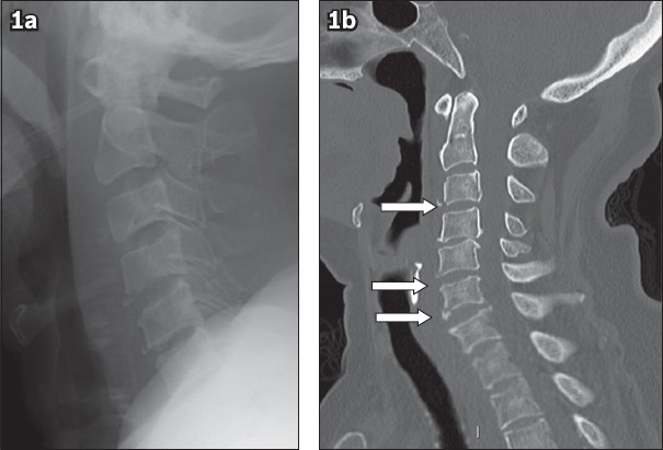
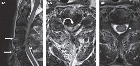
An 82-year-old woman presented with neck pain and bilateral upper limb paraesthesia after sustaining an unwitnessed fall at home the day before. Physical examination revealed tenderness over the C4-6 region but no evidence of step deformity or neurological deficit. Magnetic resonance imaging of the cervical spine revealed multiple small fractures at the anteroinferior endplate corners of the C3, C5 and C6 vertebrae with focal kyphosis and marrow oedema at these levels, as well as associated disruption of the anterior longitudinal ligament and central spinal canal stenosis. The diagnosis of multiple flexion teardrop fractures was made based on these imaging findings, and the patient subsequently received conservative management. This paper illustrates the radiological features of flexion teardrop fractures and highlights the importance of prompt diagnosis and management of such cases.
Keywords: cervical spine injury; flexion teardrop fracture; magnetic resonance imaging; vertebral fracture.
Copyright: © Singapore Medical Association.
Figures


Similar articles
-
Management of hyper-flexion injury-related teardrop fracture in an adolescent.BMJ Case Rep. 2016 Jan 28;2016:bcr2015211876. doi: 10.1136/bcr-2015-211876. BMJ Case Rep. 2016. PMID: 26822787 Free PMC article.
-
Comparison of outcomes for unstable lower cervical flexion teardrop fractures managed with halo thoracic vest versus anterior corpectomy and plating.Spine (Phila Pa 1976). 2002 Jan 15;27(2):160-6. doi: 10.1097/00007632-200201150-00008. Spine (Phila Pa 1976). 2002. PMID: 11805662
-
Progression of local kyphosis after conservative treatment for compressive cervical spine fracture with spinal cord injury.J Orthop Surg Res. 2019 Apr 11;14(1):98. doi: 10.1186/s13018-019-1115-z. J Orthop Surg Res. 2019. PMID: 30971275 Free PMC article.
-
Traumatic fracture-dislocation of C5 on C6 through a previously solid multilevel anterior cervical discectomy and fusion: a case report and review of the literature.Spine J. 2006 Jan-Feb;6(1):55-60. doi: 10.1016/j.spinee.2005.06.014. Epub 2005 Dec 6. Spine J. 2006. PMID: 16413449 Review.
-
Temporary fusionless posterior occipitocervical fixation for a proximal junctional type II odontoid fracture after previous C2-pelvis fusion: case report, description of a new surgical technique, and review of the literature.Eur Spine J. 2017 May;26(Suppl 1):243-248. doi: 10.1007/s00586-017-5093-8. Epub 2017 Apr 13. Eur Spine J. 2017. PMID: 28409288 Review.
References
-
- Kahn EA, Schneider RC. Chronic neurological sequelae of acute trauma to the spine and spinal cord. I. The significance of the acute-flexion or tear-drop fracture-dislocation of the cervical spine. J Bone Joint Surg Am. 1956;38-A:985–97. - PubMed
-
- el-Khoury GY, Kathol MH, Daniel WW. Imaging of acute injuries of the cervical spine: value of plain radiography, CT, and MR imaging. AJR Am J Roentgenol. 1995;164:43–50. - PubMed
-
- Lee C, Kim KS, Rogers LF. Triangular cervical vertebral body fractures: diagnostic significance. AJR Am J Roentgenol. 1982;138:1123–32. - PubMed
-
- Jacobs LM, Schwartz R. Prospective analysis of acute cervical spine injury: a methodology to predict injury. Ann Emerg Med. 1986;15:44–9. - PubMed
Publication types
MeSH terms
LinkOut - more resources
Full Text Sources
Medical
Miscellaneous