. 2019 Apr 15;527(6):1056-1069.
doi: 10.1002/cne.24588. Epub 2018 Dec 24.

Characterization and gonadal hormone regulation of a sexually dimorphic corticotropin-releasing factor receptor 1 cell group

Affiliations

Characterization and gonadal hormone regulation of a sexually dimorphic corticotropin-releasing factor receptor 1 cell group

Zachary J Rosinger et al. J Comp Neurol. .

Abstract

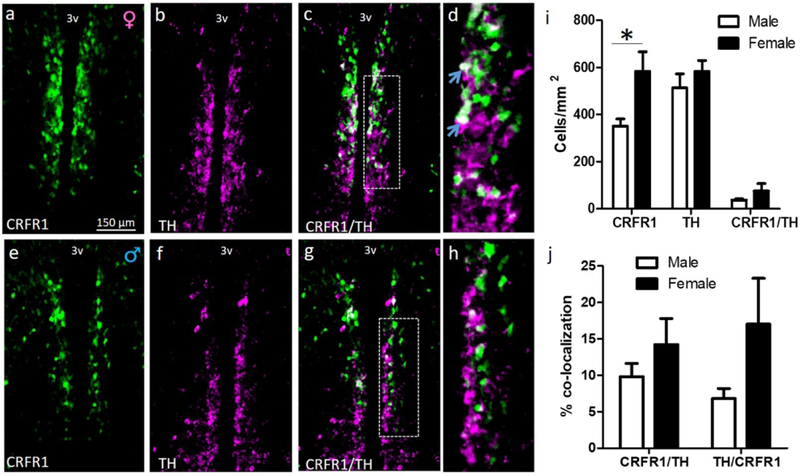
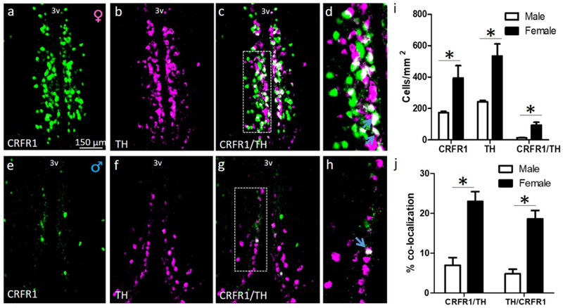
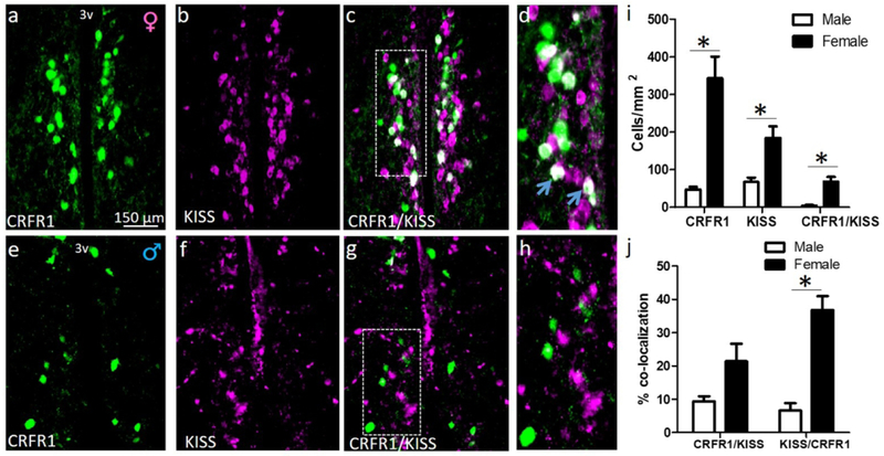
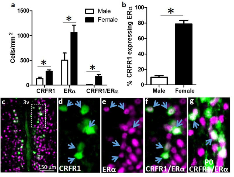
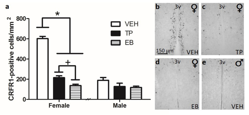
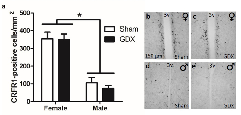
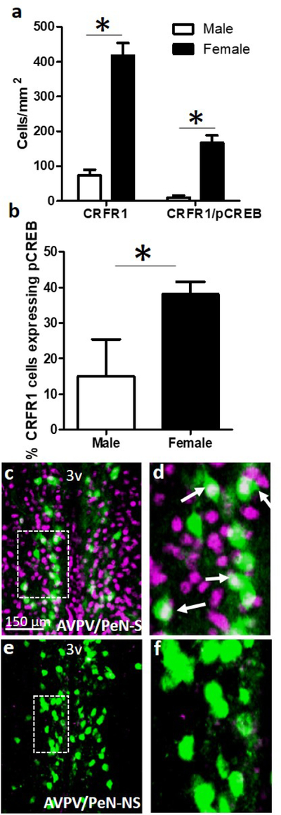
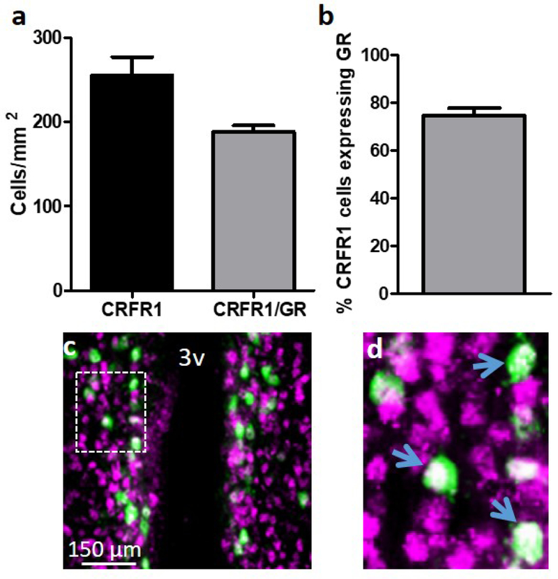
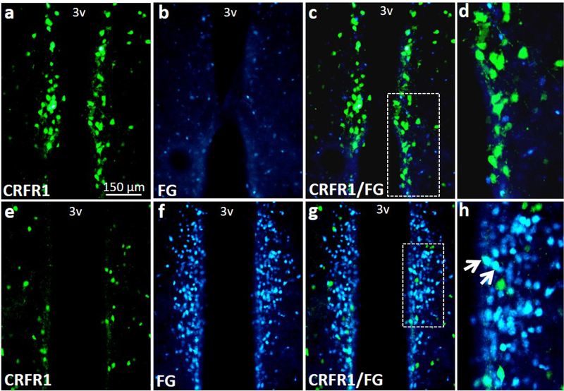
Corticotropin-releasing factor binds with high affinity to CRF receptor 1 (CRFR1) and is implicated in stress-related mood disorders such as anxiety and depression. Using a validated CRFR1-green fluorescent protein (GFP) reporter mouse, our laboratory recently discovered a nucleus of CRFR1 expressing cells that is prominent in the female rostral anteroventral periventricular nucleus (AVPV/PeN), but largely absent in males. This sex difference is present in the early postnatal period and remains dimorphic into adulthood. The present investigation sought to characterize the chemical composition and gonadal hormone regulation of these sexually dimorphic CRFR1 cells using immunohistochemical procedures. We report that CRFR1-GFP-ir cells within the female AVPV/PeN are largely distinct from other dimorphic cell populations (kisspeptin, tyrosine hydroxylase). However, CRFR1-GFP-ir cells within the AVPV/PeN highly co-express estrogen receptor alpha as well as glucocorticoid receptor. A single injection of testosterone propionate or estradiol benzoate on the day of birth completely eliminates the AVPV/PeN sex difference, whereas adult gonadectomy has no effect on CRFR1-GFP cell number. These results indicate that the AVPV/PeN CRFR1 is regulated by perinatal but not adult gonadal hormones. Finally, female AVPV/PeN CRFR1-GFP-ir cells are activated following an acute 30-min restraint stress, as assessed by co-localization of CRFR1-GFP cells with phosphorylated (p) CREB. CRFR1-GFP/pCREB cells were largely absent in the male AVPV/PeN. Together, these data indicate a stress and gonadal hormone responsive nucleus that is unique to females and may contribute to sex-specific stress responses.

Keywords: RRID: AB_2155786; RRID: AB_2296529; RRID: AB_2561044; RRID: AB_300798; RRID: AB_390204; RRID: AB_631470; anteroventral periventricular nucleus; corticotropin-releasing factor receptor 1; estrogen receptor alpha; glucocorticoid receptor; gonadal hormones; sexual differentiation.


Figures

Figure 1.
Figure 1.
Dual-label fluorescent immunohistochemistry comparing female (a-d) and male (e-h) AVPV/PeN CRFR1-GFP, tyrosine hydroxylase (TH), and co-localization of CRFR1/TH on the day of birth (d, h; high-magnification display of images c, g respectively). (i) Comparison of male versus female CRFR1-GFP- and TH-expressing cells (green, magenta, respectively), and co-expression of TH/CRFR1-GFP (white) illustrating a significant sex difference in P0 CRFR1 (F>M), but no observed differences in TH-ir. (j) Female versus male comparison of the percentage of CRFR1 cells that co-localize TH, and percentage of TH cells that co-express CRFR1. * indicates statistical significance (p≤0.05), data reported as mean ± SEM. Inset boxes indicate regions that were further magnified. Arrows indicate examples of co-localized cells. N=5 per sex. 3V, third ventricle.
Figure 2.
Figure 2.
Dual-label fluorescent immunohistochemistry comparing the adult (P60) female (a-d) and male (e-h) AVPV/PeN CRFR1-GFP, tyrosine hydroxylase (TH), and co-localization of CRFR1/TH (d, h; high-magnification display of images c, g respectively). (i) Comparison of female versus male CRFR1-GFP- (green), TH-expressing cells (magenta), and co-expression of TH in CRFR1-GFP cells (white) illustrating significant sex differences in P60 CRFR1-GFP-ir, TH-ir, and co-labeled cells (all F>M). (j) Female versus male comparison of the percentage of CRFR1 cells that co-localize TH, and percentage of TH cells that co-express CRFR1 (both F>M). * indicates statistical significance (p≤0.05), data are reported as mean ± SEM. Inset boxes indicate regions that were further magnified. Arrows indicate examples of co-localized cells. N=5 per sex. 3V, third ventricle.
Figure 3.
Figure 3.
Dual-label fluorescent immunohistochemistry comparing adult (P60) female (a-d) and male (e-h) AVPV/PeN CRFR1-GFP (a, e), kisspeptin (KISS) (b, f), and co-expression (c, g). (d, h) high magnification display of images c, g respectively. (i) Comparison of female and male CRFR1-GFP-ir and KISS-ir, with significant differences in the number of CRFR1-GFP-ir (F>M), KISS-ir (F>M), and co-localized cells (F>M). (j) Female versus male comparison of the percentage of CRFR1 cells that co-localize KISS, and percentage of KISS cells that co-express CRFR1 (both F>M). * indicates statistical significance (p≤0.05), data are reported as mean ± SEM. Inset boxes indicate regions that were further magnified. Arrows indicate examples of colocalized cells. N=5 per sex. 3V, third ventricle.
Figure 4.
Figure 4.
(a,b) Co-localization of CRFR1-GFP and ERα. Dual-label fluorescent immunohistochemistry showing that females have significantly more CRFR1-GFP, ERα, CRFR1-GFP/ERα co-localized, and a higher percentage of CRFR1-GFP cells that co-express ERα compared to males. (c-f) Representative images from a female AVPV/PeN showing a low magnification image of co-localized cells (c), CRFR1-GFP (d; green), ERα (e; magenta), and coexpression of CRFR1/ERα (f; white). (g) Image showing co-expression of CRFR1 and ERα on the day of birth (P0) in the female mouse AVPV/PeN. Inset box indicates region that was further magnified. Arrows indicate examples of co-localized cells. * Indicates statistical significance (p≤0.05), data are reported as mean ± SEM. N=5 per sex. 3V, third ventricle.
Figure 5.
Figure 5.
Neonatal (P0) hormone supplement influence on CRFR1-GFP+ cell number in (a) female versus male mice treated with sesame oil (VEH), testosterone propionate (TP), or estradiol benzoate (EB). P0 treatment with either TP or EB significantly reduced the cell number in female mice, while having no impact on the male AVPV/PeN CRFR1-GFP+ cells. Treatment with EB reduced CRFR1-GFP to a greater extent than TP. Representative images from females treated with VEH (b), TP (c), EB (d), and a male treated with VEH (e). * Indicates statistical significance (p≤0.001), + indicates p≤.05. Data are reported as mean ± SEM. N=6–7 per sex/treatment. 3V, third ventricle.
Figure 6.
Figure 6.
Adult gonadectomy effects on CRFR1-GFP+ cells. (a) Gonadectomy did not alter CRFR1-GFP+ cell number in either males or females. Representative images from female (b) and male (d) sham surgery compared with female (c) and male (e) gonadectomy, showing that removal of adult gonadal hormones does not change the expression pattern. * Indicates significant sex difference (p≤0.001), data are reported as mean ± SEM. N=8 per sex/treatment. 3V, third ventricle.
Figure 7.
Figure 7.
Dual-label fluorescent immunohistochemistry showing (a) female versus male expression levels of CRFR1-GFP (F>M), and CRFR1-GFP/pCREB colocalization (F>M). (b) Comparison between female and male co-expression, showing a sex difference in the percentage of activated CRFR1-GFP+ cells (pCREB co-expression) following a 30-minute restraint stress (F>M). Low and high magnification representative images of a female AVPV/PeN from a (c-d) s; stressed and (e-f) ns; non-stressed mouse. * Indicates statistical significance (p≤0.05), data are reported as mean ± SEM. Inset boxes indicate regions that were further magnified. Arrows indicate examples of co-localized cells. N= 5 male, 7 female mice. 3V, third ventricle.
Figure 8.
Figure 8.
Dual-label fluorescent immunohistochemistry showing the number of (a) CRFR1-GFP and CRFR1-GFP/GR co-expressing cells, and (b) the percentage of female AVPV/PeN CRFR1GFP+ cells that express GR (~75%). (c) Representative image of CRFR1 (green) and GR (magenta) co-expression (white), and (d) high magnification image of co-localized cells in the AVPV/PeN. Inset box in (c) indicates region further magnified in (d). Arrows indicate examples of co-localized cells. N= 4 female mice. 3V, third ventricle.
Figure 9.
Figure 9.
Images showing fluorogold and CRFR1-GFP labeling. Few neurosecretory (fluorogold) were found near the sexually dimorphic CRFR1 AVPV/PeN nucleus and no CRFR1GFP/fluorogold co-labeled cells were found in this region (a-d). However, high levels of fluorogold and CRFR1/fluorogold co-labeling was found in further caudal periventricular sections (e-h). Inset boxes indicate regions that were further magnified. Arrows indicate examples of colocalized cells. 3V, third ventricle.

References

    1. Bale TL, Anderson KR, Roberts AJ, Lee KF, Nagy TR, Vale WW. Corticotropin-releasing factor receptor-2-deficient mice display abnormal homeostatic responses to challenges of increased dietary fat and cold. Endocrinology 2003; 144(6):2580–2587. - PubMed
    1. Bale TL, Picetti R, Contarino A, Koob GF, Vale WW, Lee KF. Mice deficient for both corticotropin-releasing factor receptor 1 (CRFR1) and CRFR2 have an impaired stress response and display sexually dichotomous anxiety-like behavior. Journal of Neuroscience 2002; 22(1):193–199. - PMC - PubMed
    1. Bale TL, Vale WW. Increased depression-like behaviors in corticotropin-releasing factor receptor-2 deficient mice: sexually dichotomous responses. Journal of Neuroscience 2003; 23(12): 5295–5301. - PMC - PubMed
    1. Bangasser DA, Wiersielis KR, Khantsis S. Sex differences in the locus coeruleus-norepinephrine system and its regulation by stress. Brain Research 2016; 1641(Pt B):177–88. - PMC - PubMed
    1. Bealer SL, Johnson AK. Preoptic-hypothalamic periventricular lesions after food-associated drinking and circadian rhythms. Journal of Comparative Physiology and Psychology 1980; 94(3):547–55. - PubMed

Publication types

Substances