Review
. 2019 Jan;286(1):8-23.
doi: 10.1111/febs.14712. Epub 2018 Dec 18.

p62/SQSTM1: 'Jack of all trades' in health and cancer

Affiliations
Review

p62/SQSTM1: 'Jack of all trades' in health and cancer

Pablo Sánchez-Martín et al. FEBS J. 2019 Jan.

Abstract

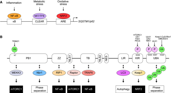
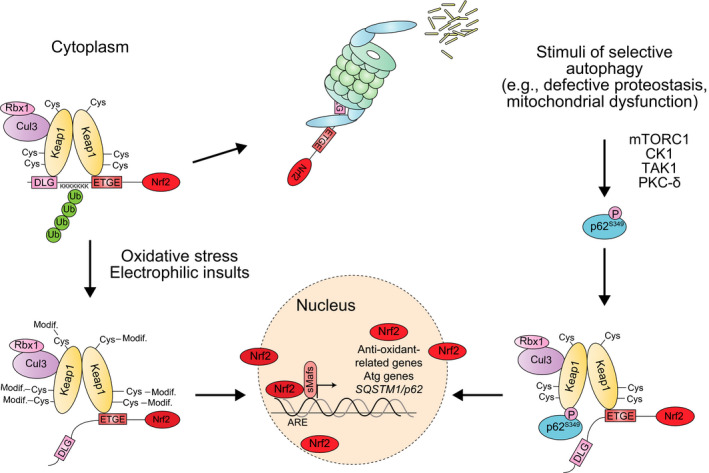
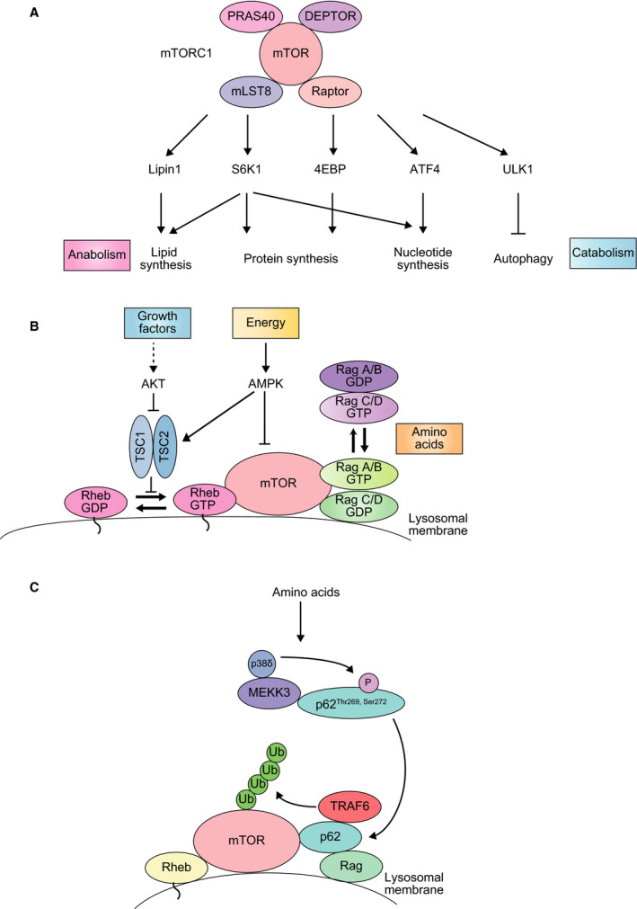
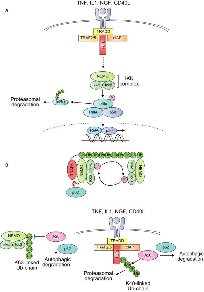
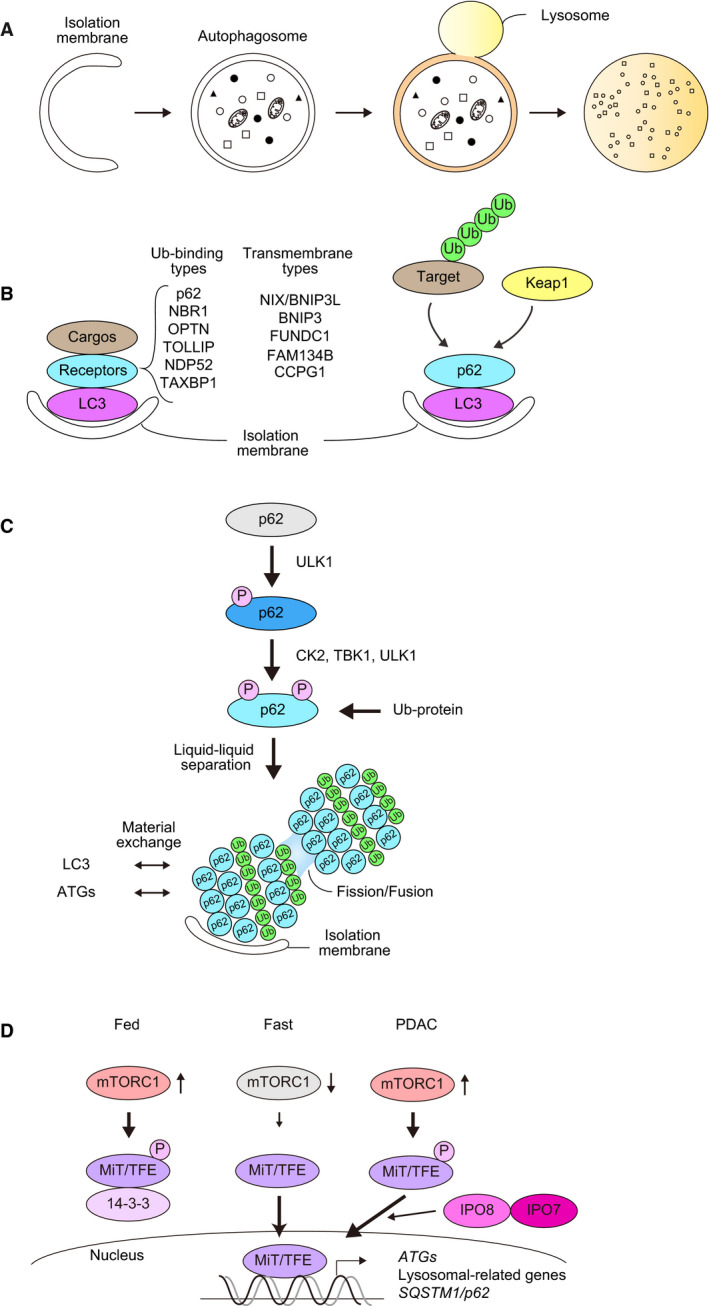
p62 is a stress-inducible protein able to change among binding partners, cellular localizations and form liquid droplet structures in a context-dependent manner. This protein is mainly defined as a cargo receptor for selective autophagy, a process that allows the degradation of detrimental and unnecessary components through the lysosome. Besides this role, its ability to interact with multiple binding partners allows p62 to act as a main regulator of the activation of the Nrf2, mTORC1, and NF-κB signaling pathways, linking p62 to the oxidative defense system, nutrient sensing, and inflammation, respectively. In the present review, we will present the molecular mechanisms behind the control p62 exerts over these pathways, their interconnection and how their deregulation contributes to cancer progression.

Keywords: Keap1; NF-κB; Nrf2; autophagy; cancer; mTORC1; p62/SQSTM1.


Figures

Figure 1
Figure 1
(A) Gene expression of SQSTM1/p62 through stress‐responsible transcription factors. CLEAR: coordinated lysosomal expression regulation. ARE: antioxidant response element. (B) Domain structure of the p62 protein. Through the interaction with multiple proteins, p62 serves as a signaling hub that modulates a variety of cellular functions (anabolism and catabolism) and/or protein property (phase separation), which are regulated by post‐translational modifications of p62. PB1: Phox and Bem1p. ZZ: Zinc finger. NLS: nuclear localization signal. TB: TRAF6‐binding domain. NES: nuclear export signal. LIR: LC3‐interacting region. KIR: Keap1‐interacting region. UBA: ubiquitin‐associated.
Figure 2
Figure 2
Mechanism of Nrf2 activation dependent on S349 phosphorylation of p62. Left: under basal conditions, Keap1, in collaboration with the Cul3/Rbx1 E3 ubiquitin ligase complex, promotes the proteasomal degradation of Nrf2. Upon oxidative stress, Keap1 oxidation results in the liberation of Nrf2 and its translocation to the nucleus, where it promotes the expression of a battery of target genes encoding antioxidant proteins and anti‐inflammatory enzymes. Right: phosphorylated p62 interacts with the Nrf2‐binding site of Keap1 and competitively inhibits the Keap1–Nrf2 interaction, resulting in the expression of Nrf2 target genes, which include p62. Therefore, Ser349 phosphorylated p62 causes the constitutive activation of Nrf2.
Figure 3
Figure 3
(A) The mTORC1 pathway. The mTORC1 complex is formed by the mTOR kinase, the regulator subunits Raptor and PRAS40 and the inhibitors mLST8 and DEPTOR. Upon mTORC1 stimulation, downstream effectors promote anabolism and suppress catabolism. (B) The activity of mTORC1 is inhibited by the TSC complex, which is itself inactivated in the presence of growth factors and upregulated upon energy deprivation. In the later situation, AMPK inhibits mTORC1 both directly and through TSC activation. Increased amino acid availability converts RagA/B GDP and RagC/D GTP into RagA/B GTP and RagC/D GDP respectively, assembling mTORC1 on the lysosome, where Rheb activates mTORC1. (C) mTORC1 activation by p62. Amino acid stimulation promotes the phosphorylation of p62 at Thr269 and Ser272 by MEKK3/p38delta. This results in the formation of a signaling hub over the lysosomal membrane through the interaction of p62 with Raptor, TRAF6 and Rag proteins. As a consequence, mTOR is ubiquitinated by TRAF6, and then activated by Rheb.
Figure 4
Figure 4
(A) The NF‐κB pathway. Multiple signals, such as proinflammatory cytokines or growth factors, can induce NF‐κB activation. Following the ligand‐receptor binding, the activation of the IKK complex releases NF‐κB (RelA–p50 complex) from its inhibitor IκB. This is achieved through the phosphorylation and subsequent proteasomal degradation of the inhibitor. Then, NF‐κB translocates to the nucleus and induces the expression of target genes, including p62. (B) Two mechanisms of p62‐mediated NF‐κB activation. First, p62 seems to be required for NEMO ubiquitination by TRAF6. This ubiquitination is recognized by the NEMO subunits of other IKK complexes, bringing them into the close proximity required for their cross‐phosphorylation and activation. Second, p62 promotes the autophagic degradation of the ubiquitin‐editing enzyme A20, which otherwise inhibits NF‐κB activation by promoting RIP1 degradation and reversing NEMO ubiquitination.
Figure 5
Figure 5
(A) Macroautophagy is accompanied by dynamic membrane biogenesis and autophagosome formation. The autophagosome sequesters a portion of the cytoplasm and fuses with the lysosome, where its contents are degraded. (B) Receptor‐mediated selective autophagy. Receptor proteins are divided into two groups: ubiquitin‐binding type and transmembrane type. Both have the ability to bind to Atg8 family proteins (e.g., LC3s and GABARAPs). p62 is a representative Ub‐binding type receptor, and it also mediates autophagic degradation of certain proteins (e.g., Keap1) through their direct interaction without prior ubiquitination. (C) p62‐mediated selective autophagy. When selective autophagic cargos such as misfolded proteins appear in the cytoplasm, they are ubiquitinated. Concomitantly, Ser407, located at the UBA domain of p62, is initially phosphorylated by ULK1 kinase. This phosphorylation destabilizes the UBA dimer interface and subsequently casein kinase 2 (CK2), TANK‐binding kinase 1 (TBK1), or ULK1 phosphorylate Ser403 of the UBA domain, which increases the binding affinity of p62 for the ubiquitin chain. The p62 forming a complex with ubiquitin chains has liquid‐like properties and forms droplets in which LC3 and other Atg proteins assemble to form the isolation membrane. Keap1, a binding protein for p62 may also shuttle from cytoplasm to the droplets. (D) Regulation of the nuclear translocation of the MiT‐TFE family by mTORC1‐mediated phosphorylation. Under nutrient‐rich conditions, the MiT‐TFE family is phosphorylated by mTORC1 and subsequently trapped by 14‐3‐3 protein. As a result, the MiT‐TFE family is kept in the cytoplasm. Once mTORC1 is inactivated by metabolic stresses such as nutrient deprivation, the MiT‐TFE family translocate into the nucleus to induce lysosomal biogenesis, Atg genes and p62/SQSTM1. In PDAC, increased level of Importin8 and 7 bypasses the regulation of mTORC1.

References

    1. Svenning S, Lamark T, Krause K & Johansen T (2011) Plant NBR1 is a selective autophagy substrate and a functional hybrid of the mammalian autophagic adapters NBR1 and p62/SQSTM1. Autophagy 7, 993–1010. - PMC - PubMed
    1. Ling J, Kang Y, Zhao R, Xia Q, Lee DF, Chang Z, Li J, Peng B, Fleming JB, Wang H et al (2012) KrasG12D‐induced IKK2/beta/NF‐kappaB activation by IL‐1alpha and p62 feedforward loops is required for development of pancreatic ductal adenocarcinoma. Cancer Cell 21, 105–120. - PMC - PubMed
    1. Jain A, Lamark T, Sjottem E, Larsen KB, Awuh JA, Overvatn A, McMahon M, Hayes JD & Johansen T (2010) p62/SQSTM1 is a target gene for transcription factor NRF2 and creates a positive feedback loop by inducing antioxidant response element‐driven gene transcription. J Biol Chem 285, 22576–22591. - PMC - PubMed
    1. Settembre C, Di Malta C, Polito VA, Garcia Arencibia M, Vetrini F, Erdin S, Erdin SU, Huynh T, Medina D, Colella P et al (2011) TFEB links autophagy to lysosomal biogenesis. Science 332, 1429–1433. - PMC - PubMed
    1. Rea SL, Majcher V, Searle MS & Layfield R (2014) SQSTM1 mutations–bridging Paget disease of bone and ALS/FTLD. Exp Cell Res 325, 27–37. - PubMed

Publication types