

Estrogen but not testosterone preserves myofilament function from doxorubicin-induced cardiotoxicity by reducing oxidative modifications
- PMID: 30499711
- PMCID: PMC6397386
- DOI: 10.1152/ajpheart.00428.2018
Estrogen but not testosterone preserves myofilament function from doxorubicin-induced cardiotoxicity by reducing oxidative modifications
Abstract
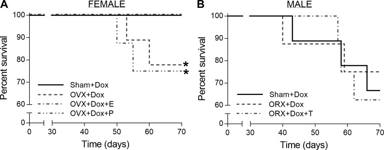
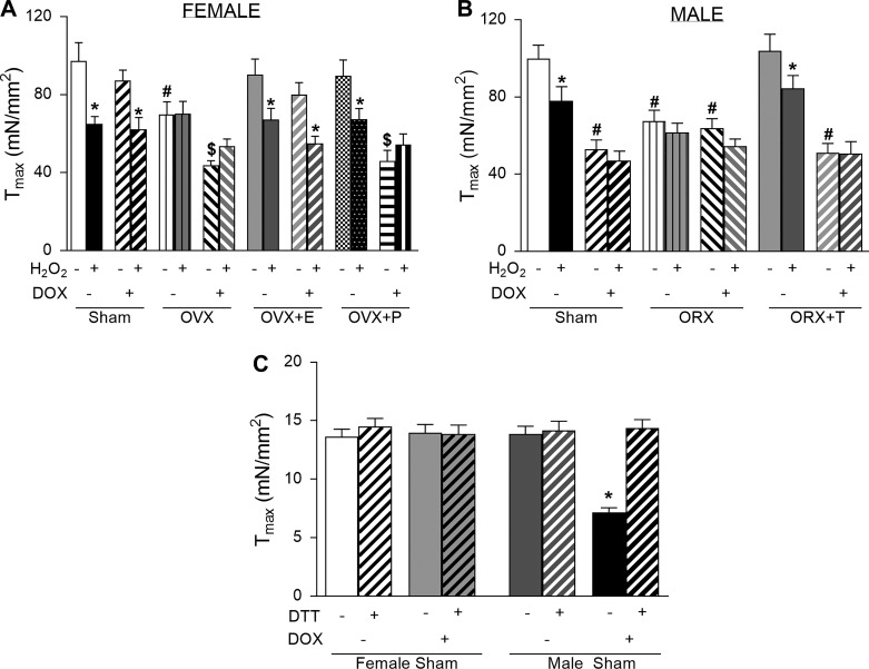
Here, we aimed to explore sex differences and the impact of sex hormones on cardiac contractile properties in doxorubicin (DOX)-induced cardiotoxicity. Male and female Sprague-Dawley rats were subjected to sham surgery or gonadectomy and then treated or untreated with DOX (2 mg/kg) every other week for 10 wk. Estrogen preserved maximum active tension (Tmax) with DOX exposure, whereas progesterone and testosterone did not. The effects of sex hormones and DOX correlated with both altered myosin heavy chain isoform expression and myofilament protein oxidation, suggesting both as possible mechanisms. However, acute treatment with oxidative stress (H2O2) or a reducing agent (DTT) indicated that the effects on Tmax were mediated by reversible myofilament oxidative modifications and not only changes in myosin heavy chain isoforms. There were also sex differences in the DOX impact on myofilament Ca2+ sensitivity. DOX increased Ca2+ sensitivity in male rats only in the absence of testosterone and in female rats only in the presence of estrogen. Conversely, DOX decreased Ca2+ sensitivity in female rats in the absence of estrogen. In most instances, this mechanism was through altered phosphorylation of troponin I at Ser23/Ser24. However, there was an additional DOX-induced, estrogen-dependent, irreversible (by DTT) mechanism that altered Ca2+ sensitivity. Our data demonstrate sex differences in cardiac contractile responses to chronic DOX treatment. We conclude that estrogen protects against chronic DOX treatment in the heart, preserving myofilament function. NEW & NOTEWORTHY We identified sex differences in cardiotoxic effects of chronic doxorubicin (DOX) exposure on myofilament function. Estrogen, but not testosterone, decreases DOX-induced oxidative modifications on myofilaments to preserve maximum active tension. In rats, DOX exposure increased Ca2+ sensitivity in the presence of estrogen but decreased Ca2+ sensitivity in the absence of estrogen. In male rats, the DOX-induced shift in Ca2+ sensitivity involved troponin I phosphorylation; in female rats, this was through an estrogen-dependent mechanism.
Conflict of interest statement
No conflicts of interest, financial or otherwise, are declared by the authors.
Figures


Similar articles
-
Rapid changes in cardiac myofilament function following the acute activation of estrogen receptor-alpha.PLoS One. 2012;7(7):e41076. doi: 10.1371/journal.pone.0041076. Epub 2012 Jul 30. PLoS One. 2012. PMID: 22859967 Free PMC article.
-
Rat cardiac troponin T mutation (F72L)-mediated impact on thin filament cooperativity is divergently modulated by α- and β-myosin heavy chain isoforms.Am J Physiol Heart Circ Physiol. 2015 Oct;309(8):H1260-70. doi: 10.1152/ajpheart.00519.2015. Epub 2015 Sep 4. Am J Physiol Heart Circ Physiol. 2015. PMID: 26342069 Free PMC article.
-
Sexually dimorphic myofilament function and cardiac troponin I phosphospecies distribution in hypertrophic cardiomyopathy mice.Arch Biochem Biophys. 2013 Jul 1;535(1):39-48. doi: 10.1016/j.abb.2012.12.023. Epub 2013 Jan 23. Arch Biochem Biophys. 2013. PMID: 23352598 Free PMC article.
-
Oxidation of cardiac myofilament proteins: Priming for dysfunction?Mol Aspects Med. 2018 Oct;63:47-58. doi: 10.1016/j.mam.2018.08.003. Epub 2018 Aug 23. Mol Aspects Med. 2018. PMID: 30130564 Review.
-
A comprehensive review on the effects of sex hormones on chemotherapy-induced cardiotoxicity: are they lucrative or unprofitable?Cardiooncology. 2024 Dec 3;10(1):86. doi: 10.1186/s40959-024-00293-3. Cardiooncology. 2024. PMID: 39627907 Free PMC article. Review.
Cited by
-
Advantages of prophylactic versus conventionally scheduled heart failure therapy in an experimental model of doxorubicin-induced cardiomyopathy.J Transl Med. 2019 Jul 19;17(1):229. doi: 10.1186/s12967-019-1978-0. J Transl Med. 2019. PMID: 31324258 Free PMC article.
-
2-Methoxyestradiol ameliorates doxorubicin-induced cardiotoxicity by regulating the expression of GLUT4 and CPT-1B in female rats.Naunyn Schmiedebergs Arch Pharmacol. 2024 Sep;397(9):7129-7139. doi: 10.1007/s00210-024-03073-z. Epub 2024 Apr 23. Naunyn Schmiedebergs Arch Pharmacol. 2024. PMID: 38652282 Free PMC article.
-
The Contractile Function of Ventricular Cardiomyocytes Is More Sensitive to Acute 17β-Estradiol Treatment Compared to Atrial Cardiomyocytes.Cells. 2025 Apr 8;14(8):561. doi: 10.3390/cells14080561. Cells. 2025. PMID: 40277887 Free PMC article.
-
Doxorubicin-induced cardiotoxicity and risk factors.Int J Cardiol Heart Vasc. 2023 Dec 27;50:101332. doi: 10.1016/j.ijcha.2023.101332. eCollection 2024 Feb. Int J Cardiol Heart Vasc. 2023. PMID: 38222069 Free PMC article.
-
Voluntary wheel running reduces tumor growth and increases capillarity in the heart during doxorubicin chemotherapy in a murine model of breast cancer.Front Physiol. 2024 Apr 25;15:1347347. doi: 10.3389/fphys.2024.1347347. eCollection 2024. Front Physiol. 2024. PMID: 38725573 Free PMC article.
References
Publication types
MeSH terms
Substances
Grants and funding
LinkOut - more resources
Full Text Sources
Medical
Miscellaneous