

MRI of THA Correlates With Implant Wear and Tissue Reactions: A Cross-sectional Study
- PMID: 30499779
- PMCID: PMC6345304
- DOI: 10.1097/CORR.0000000000000535
MRI of THA Correlates With Implant Wear and Tissue Reactions: A Cross-sectional Study
Abstract
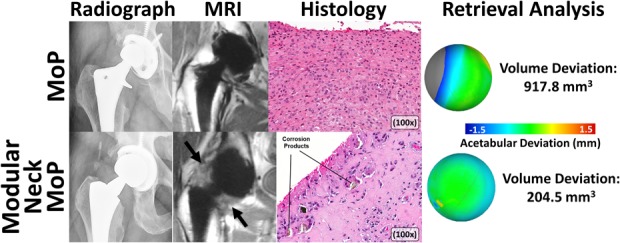
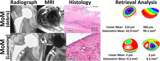
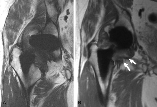
Background: MRI is predictive of adverse local tissue reactions (ALTRs) after THA but how MRI directly relates to implant surface wear, fretting, and trunnion corrosion at different articulations between implant components remains unclear. MRI generates high-contrast images to display soft tissues around arthroplasty and may provide a surgeon the means to distinguish and differentiate host-related synovial patterns as a response to either polyethylene wear or metal wear and corrosion products.
Questions/purposes: The purposes of this study were (1) to correlate findings from MRI in patients who have undergone THA with direct assessment of implant wear, corrosion, and fretting from retrieved components; and (2) to distinguish the unique synovial responses on MRI in patients who have undergone THA based on bearing materials.
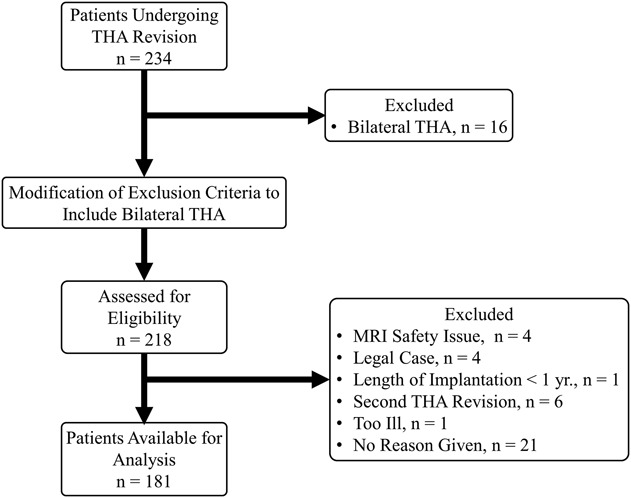
Methods: In this prospective study, patients undergoing THA (181 patients, 187 hips) with metal-on-metal (MoM), hip resurfacing (HRA), metal-on-polyethylene (MoP), ceramic-on-polyethylene, ceramic-on-ceramic, or modular neck designs having revision surgery (between October 2013 and June 2017) underwent preoperative MRI. A single reader blinded to the bearing surface made an assessment of the synovial response (Gwet's AC1, 0.65-0.97); these data were compared with semiquantitative histology of tissue samples by a single reader (Gwet's AC1, 0.92) and semiquantitative wear, corrosion, and fretting analysis of retrieved components using Goldberg scoring (Gwet's AC1, 0.60-0.79). Direct noncontact measurements of implant wear were also made. Correlations and analyses of variance were used to assess associations between metrics and differences by implant type, respectively.
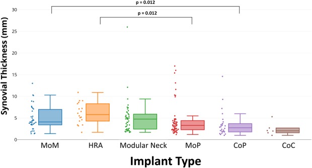
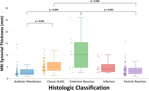
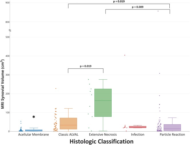
Results: Correlations were found between MRI synovial thickness with severity of fretting and corrosion damage of the female head-neck trunnion of femoral stems in modular designs (ρ = 0.26 [95% confidence interval {CI}, 0.12-0.39]; p = 0.015, n = 185) and ALTR grade and volumetric wear in MoM bearings (ρ = 0.93 [95% CI, 0.72-0.98]; p < 0.001, n = 10). MRI synovial thickness was highest in patients identified with aseptic lymphocyte-dominated vasculitis-associated lesions and diffuse tissue necrosis. On MRI, MoP hips demonstrated a distinct polymeric synovial response, whereas HRA, MoM, and modular hips more commonly demonstrated ALTR. Hips classified as having a polymeric synovial response on MRI had a greater number of particles present in tissue samples.
Conclusions: In this study, we demonstrated that MRI of THA can distinguish synovial responses that reflect the bearing type of the implanted THA and correlate to direct measurements of implant wear, corrosion, and fretting and histologic assessment of wear particles in periprosthetic tissues. MRI provides a means of direct, noninvasive visualization of the host-generated synovial response. Patients presenting with painful arthroplasties may be evaluated for the cause of their discomfort, specifically highlighting any concerning synovial reactions that would warrant more prompt surgical intervention. Future studies would benefit from a prospective evaluation of different implants to assess the natural longitudinal history of arthroplasty complications, including the development and prevalence of ALTR across bearing constructs.
Level of evidence: Level III, diagnostic study.
Conflict of interest statement
All ICMJE Conflict of Interest Forms for authors and
Figures
Comment in
-
CORR Insights®: MRI of THA Correlates With Implant Wear and Tissue Reactions: A Cross-sectional Study.Clin Orthop Relat Res. 2019 Jan;477(1):175-176. doi: 10.1097/CORR.0000000000000582. Clin Orthop Relat Res. 2019. PMID: 30499781 Free PMC article. No abstract available.
References
-
- Amstutz HC, Grigoris P. Metal on metal bearings in hip arthroplasty. Clin Orthop Relat Res. 1996;329(Suppl):S11-34. - PubMed
-
- Anderson H, Toms AP, Cahir JG, Goodwin RW, Wimhurst J, Nolan JF. Grading the severity of soft tissue changes associated with MOM hip replacements: reliability of an MR grading system. Skeletal Radiol. 2011;40:303-307. - PubMed
-
- Barlow BT, Boles JW, Lee YY, Ortiz PA, Westrich GH. Short-term outcomes and complications after rejuvenate modular total hip arthroplasty revision. J Arthroplasty. 2016;31:857-862. - PubMed
Publication types
MeSH terms
Grants and funding
LinkOut - more resources
Full Text Sources
Medical
Research Materials
