. 2018 Nov;66(11):2503-2513.
doi: 10.1002/glia.23501.

Activated T cells induce proliferation of oligodendrocyte progenitor cells via release of vascular endothelial cell growth factor-A

Affiliations

Activated T cells induce proliferation of oligodendrocyte progenitor cells via release of vascular endothelial cell growth factor-A

Elliot H Choi et al. Glia. 2018 Nov.

Abstract

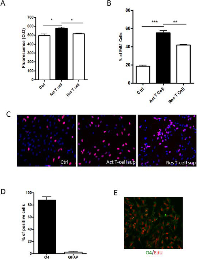
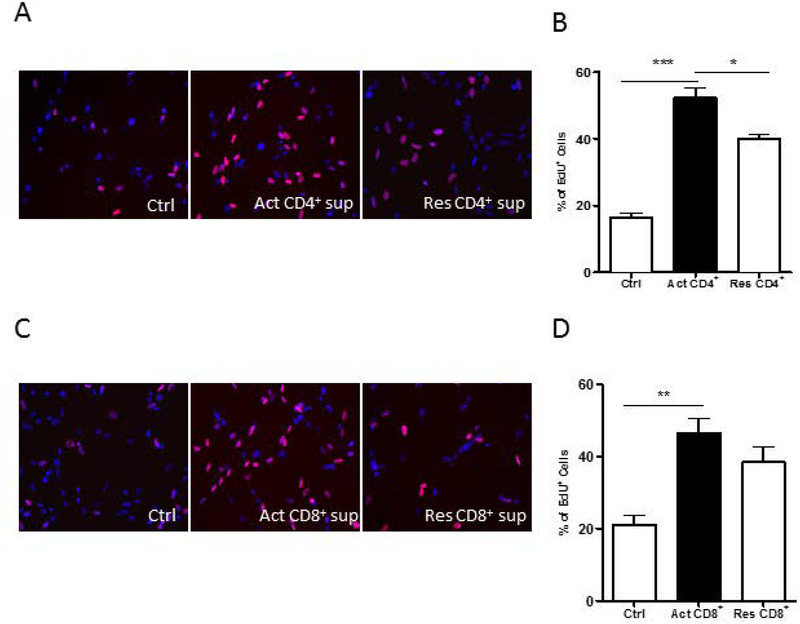
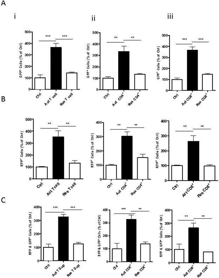
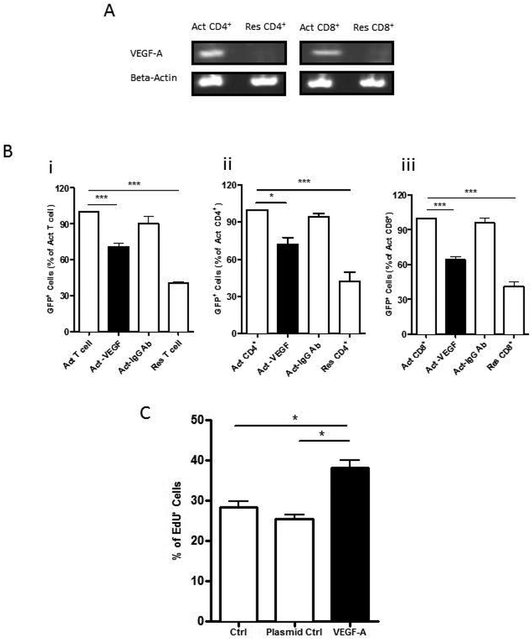
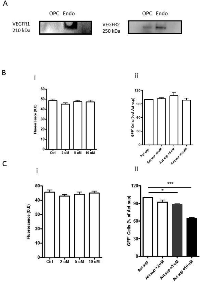
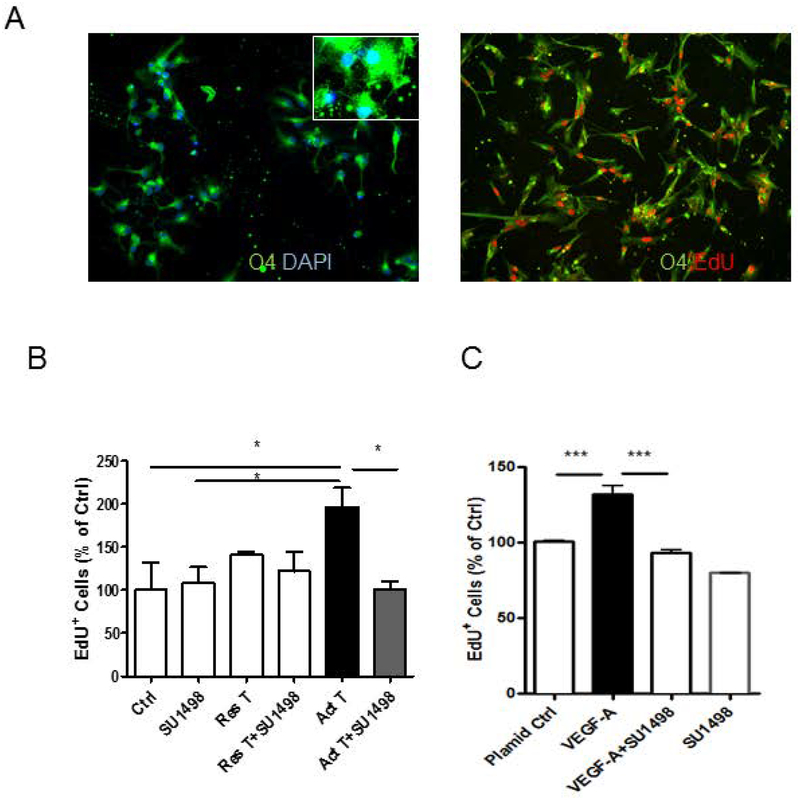
Neuroinflammatory diseases such as multiple sclerosis are characterized by infiltration of lymphocytes into the central nervous system followed by demyelination and axonal degeneration. While evidence suggests that activated T lymphocytes induce neurotoxicity and impair function of neural stem cells, the effect of T cells on oligodendrocyte progenitor cells (OPCs) is still uncertain, partly due to the difficulty in obtaining human OPCs. Here we studied the effect of activated T cells on OPCs using OPCs derived from human hematopoietic stem cells or from human fetal brain. OPCs were exposed to supernatants (sups) from activated T cells. Cell proliferation was determined by EdU incorporation and CellQuanti-Blue assays. Surprisingly, we found that sups from activated T cells induced OPC proliferation by regulating cell cycle progression. Vascular endothelial growth factor A (VEGF-A) transcripts were increased in T cells after activation. Immunodepletion of VEGF-A from activated T cell sups significantly attenuated its effect on OPC proliferation. Furthermore, VEGF receptor 2 (VEGFR2) was expressed on OPCs and its inhibition also attenuated activated T cell-induced OPC proliferation. Thus, activated T cells have a trophic role by promoting OPC proliferation via the VEGFR2 pathway.

Keywords: T cells; VEGF; neural inflammation; neural stem cells; oligodendrocyte progenitor cells.


Conflict of interest statement

Conflict of Interest: The authors declare no competing financial interests.

Figures

Figure 1:
Figure 1:. Effect of T-cell sups on OPCs.
(A) OPCs were exposed to culture sups from T cells for 24 hrs. An increase in the number of OPCs was seen with activated (Act) T-cell sups compared to T-cell media control (Ctrl) or resting (Res) T-cell sup as determined by CellQuanti-blue assay. (B) An increase in the percentage of proliferating cells is seen with Act T sup as determined by uptake of EdU. (C) Representative photomicrographs show EdU staining (red) OPCs exposed to T-cell media Ctrl, Act T-cell sup or Res T-cell sup. The nuclei are stained blue with DAPI. (D) OPCs exposed to Act T-cell sup were dual stained for O4 or GFAP and EdU. While most of the cells were positive for O4 and only a few were positive for GFAP, nearly all proliferating cells were O4 positive. (E) Representative photomicrograph shows O4 cells (green) dual stained for EdU (red). Data represent mean + SEM of three independent experiments. N=3 for A, B and D, *P<0.05; **P<0.005 and ***P<0.0005.
Figure 2:
Figure 2:. Effect of CD4 and C8 lymphocytes on OPC proliferation.
(A) Representative photomicrographs of OPCs exposed to T cell media control (Ctrl), activated (Act) CD4+ cell sups or resting (Res) CD4+ cell sups. Nuclei are stained blue with DAPI and EdU+ cells are red. (B) Act -CD4+ cell sups induced increased proliferation of OPCs as determined by percentage of EdU positive cells. (C) Representative photomicrographs of OPCs exposed to Ctrl, Act CD8+ cell sup or Res CD4+ cell sup. Nuclei are stained blue with DAPI and EdU+ cells are red. (D) Act -CD8+ cell sups induced increased proliferation of the OPCs. Data represent mean + SEM of three independent experiments. N=3 for Band D, *P<0.05; **P<0.005 and ***P<0.0005
Figure 3:
Figure 3:. Effect of T cells on OPC cell cycle phases:
FUCCI Cell Cycle Sensor was used to study the cell cycle phases. (A) GFP indicates number of OPCs in S/G2/M phase. A significant GFP increase in these cells is seen with treatment with sups from activated (Act) (i) pan T cells (ii) CD4 cells or (iii) CD8 cells compared to T cell media control (Ctrl) or resting (Res) pan T cells, CD4 or CD8 cells respectively. (B) RFP+ cells indicate number of OPCs in G1 phase and (C) dual GFP and RFP+ cells indicate cells in transition phase. Data represent mean + SEM of three independent experiments. N=3, *P<0.05; **P<0.005 and ***P<0.0005
Figure 4:
Figure 4:. Effect of VEGF on OPC proliferation.
(A) Expression of VEGF-A transcripts as determined by RT-PCR is increased in activated (Act) CD4+ and CD8+ cells. (B) Fluorescence ubiquitination cell cycle indicator was used to measure OPC proliferation and is indicated by GFP+ cells. Exposure to sups following immunodepletion of VEGF (Act-VEGF) from (i) Act Pan T cells (ii) Act CD4+ cells or (iii) Act CD8+ cells resulted in a significant decrease in proliferation of OPCs compared to each of the sups treated with an isotype antibody (Act-IgG Ab) or untreated sups from each of the Act cell types. (C) OPC proliferation was determined by EdU incorporation. Sups from 293 cells transfected with VEGF-A plasmid show increased proliferation compared to sups from cells treated with transfection reagent alone (Ctrl) or transfected with a control plasmid (Plasmid Ctrl). Data represent mean + SEM from three independent experiments. N=3 for B and C, *P<0.05; ***P<0.0005
Figure 5:
Figure 5:. Effect of Activated T cell sups on OPCs is mediated by VEGFR-2.
(A) Western blot analysis for VEGF receptors shows lack of detection of VEGFR-1 but presence of VEGFR-2 on OPCs. Endothelial cells (Endo) were used as a positive control. Propidium iodide staining was used to measure cell viability and fluorescence ubiquitination cell cycle indicator was used to measure OPC proliferation and is indicated by GFP+ cells. (B) VEGFR-1 antagonist, ZM306416 (i) had no effect on OPC viability when incubated alone and (ii) did not inhibit the effect of activated (Act) T cell sup induced GFP+ cell numbers on OPCs. (C) VEGFR-2 antagonist, SU1498, (i) had no effect on OPC viability when incubated alone but (ii) inhibited OPC proliferation as indicated by GFP+ cell numbers in a dose-responsive manner in the presence of Act T cell sup. Data represent mean + SEM from three independent experiments. N=3 for B and C, **P<0.005; ***P<0.0005
Figure 6.
Figure 6.. Effect of VEGF-A on proliferation of primary human fetal OPCs.
Immunostaining for O4 was used for the identification of primary human OPCs. EdU incorporation assay was used to measure OPC proliferation. (A), almost all EdU positive cells were O4 positive cells. (B), OPCs treated with sup from activated T cells (Act T) show increased proliferation compared to control cells (Ctrl) while OPCs treated with sup from restive T cells (Res T) show no increased proliferation. The increased proliferation of OPCs was attenuated by VEGF-A inhibitor SU1498. (C), Sups from 293 cells transfected with VEGF-A plasmid show increased proliferation compared to sups from cells transfected with a control plasmid (Plasmid Ctrl), but was attenuated by VEGF-A inhibitor SU1498. Data represent mean ± SEM from three independent experiments. N=3 for B and C, *P<0.05, ***P<0.001

References

    1. Al Heialy S, Risse PA, Zeroual MA, Roman HN, Tsuchiya K, Siddiqui S, Laporte SA, Martin JG. 2013. T cell-induced airway smooth muscle cell proliferation via the epidermal growth factor receptor. Am J Respir Cell Mol Biol 49:563–70. - PubMed
    1. Argaw AT, Asp L, Zhang J, Navrazhina K, Pham T, Mariani JN, Mahase S, Dutta DJ, Seto J, Kramer EG and et al. 2012. Astrocyte-derived VEGF-A drives blood-brain barrier disruption in CNS inflammatory disease. J Clin Invest 122:2454–68. - PMC - PubMed
    1. Blackburn D, Sargsyan S, Monk PN, Shaw PJ. 2009. Astrocyte function and role in motor neuron disease: A future therapeutic target? Glia 57:1251–1264. - PubMed
    1. Chandran S, Compston A, Jauniaux E, Gilson J, Blakemore W, Svendsen C. 2004. Differential generation of oligodendrocytes from human and rodent embryonic spinal cord neural precursors. Glia 47:314–324. - PubMed
    1. Chang A, Tourtellotte WW, Rudick R, Trapp BD. 2002. Premyelinating oligodendrocytes in chronic lesions of multiple sclerosis. N Engl J Med 346:165–73. - PubMed

Publication types

MeSH terms

Substances