. 2018 Nov 29;175(6):1481-1491.e13.
doi: 10.1016/j.cell.2018.10.057.

Liquid Nuclear Condensates Mechanically Sense and Restructure the Genome

Affiliations

Liquid Nuclear Condensates Mechanically Sense and Restructure the Genome

Yongdae Shin et al. Cell. .

Erratum in

Abstract

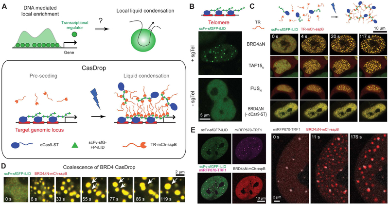
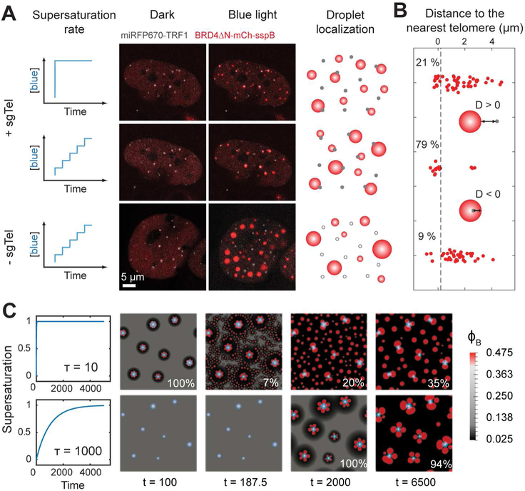
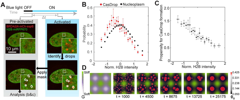
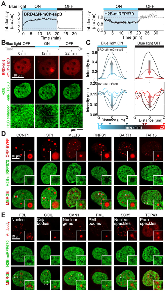
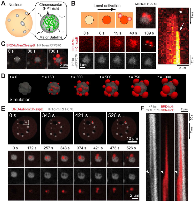
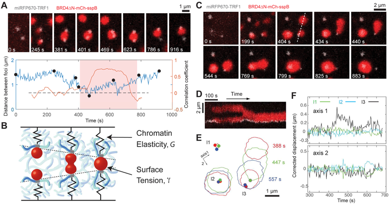
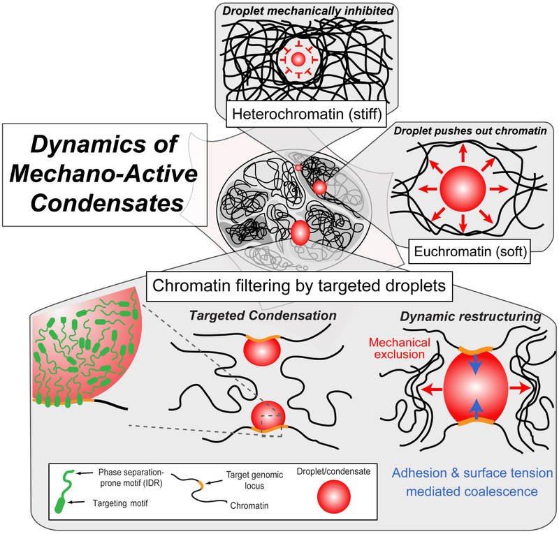
Phase transitions involving biomolecular liquids are a fundamental mechanism underlying intracellular organization. In the cell nucleus, liquid-liquid phase separation of intrinsically disordered proteins (IDPs) is implicated in assembly of the nucleolus, as well as transcriptional clusters, and other nuclear bodies. However, it remains unclear whether and how physical forces associated with nucleation, growth, and wetting of liquid condensates can directly restructure chromatin. Here, we use CasDrop, a novel CRISPR-Cas9-based optogenetic technology, to show that various IDPs phase separate into liquid condensates that mechanically exclude chromatin as they grow and preferentially form in low-density, largely euchromatic regions. A minimal physical model explains how this stiffness sensitivity arises from lower mechanical energy associated with deforming softer genomic regions. Targeted genomic loci can nonetheless be mechanically pulled together through surface tension-driven coalescence. Nuclear condensates may thus function as mechano-active chromatin filters, physically pulling in targeted genomic loci while pushing out non-targeted regions of the neighboring genome. VIDEO ABSTRACT.

Keywords: chromatin; condensates; gene regulation; mechanobiology; nuclear mechanics; nuclear organization; optogenetics; phase immiscibility; phase separation.


Conflict of interest statement

Declaration of Interests

A patent application describing the CasDrop technology is currently pending.

Figures

Figure 1.
Figure 1.
The CasDrop system enables liquid condensation of transcriptional regulators at target loci. (A) Transcriptional regulators (TRs) are often enriched at regulatory DNA elements near genes and may facilitate local liquid-liquid phase separation. The CasDrop system is designed to function similarly yet with added genome-targeting programmability and optogenetic controllability. The modular components of the CasDrop include dCas9-ST, scFv-sfGFP-iLID and TR-mch-sspB. (B) Pre-seeding at telomeres in a HEK293 cell by expressing dCas9-ST, scFv-sfGFP-iLID and sgRNA for telomeres. In the absence of the telomere-targeting sgRNA, punctate fluorescence signals are not observed. (C) HEK293 cells expressing dCas9-ST, scFv-sfGFP-iLID and mch-sspB fused to different transcriptional regulators (TR) are activated with blue light. A control without expressing the dCas9-ST scaffold shows no clustering. (D) Time lapse images of BRD4 CasDrop in a HEK293 cell showing rapid condensation followed by frequent coalescence between droplets (white arrowheads). (E) (Left) Fluorescence images of a NIH3T3 cell expressing dCas9-ST, scFv-sfGFP-iLID, BRD4ΔN-mCh-sspB, miRFP670-TRF1 and sgRNA for telomeres. Images show localization patterns of each construct prior to blue light activation. (Right) Time lapse images of the same cell during blue light illumination. See also Figure S1
Figure 2.
Figure 2.
The effects of pre-seeding and activation protocol on the localization pattern of BRD4 CasDrop. (A) Two different activation protocols are applied to the same NIH3T3 cell expressing dCas9-ST, scFv-sfGFP-iLID, BRD4ΔN-mCh-sspB, miRFP670-TRF1 and sgRNA for telomeres. Fluorescence images of the cell before and after activation protocols are shown. When the ramping protocol is applied on a cell without telomere-targeting sgRNA, assembled droplets exhibited apparently random droplet localization irrespective of telomeres. (B) For each BRD4 droplet, a distance from the droplet boundary to the nearest telomere is measured. Each red dot represents a single droplet. Fractions of droplets whose distances to the nearest telomere are smaller than 0.2 μm (black dashed line) are given in percentage. (C) Simulations of our mechanical droplet exclusion model demonstrating that during CasDrop nucleation and growth in the presence of pre-seed sites (white circles denote stiff seed cores, light blue halos denote surrounding regions of enhanced concentration, φA) targeted droplet localization improves with decreasing activation rate. Rapid and slow activation protocols are shown in the upper and lower rows, respectively, with time increasing from left to right. Fractions of droplets overlapping with a pre-seed site at the final time shown are given in percentage. See also Supplementary Videos 1-2
Figure 3.
Figure 3.
IDR-driven condensates prefer growing at regions of low chromatin density. (A) Schematic of experiment and analysis for (b) and (c). U2OS cells expressing dCas9-ST, scFv-sfGFP-iLID, BRD4ΔN-mCh-sspB, and H2B-miRFP670 are activated with blue light. Clusters of CasDrop are identified at various time (Δt = 3 to 30 s after activation starts) using mCh fluorescence signal. The resulting binary mask is then applied to pre-activation (0 s) image marking pixels where droplets will form; H2B intensity of these pixels is analyzed in (B) and (C). (B) Probability distribution of normalized H2B intensity for nucleoplasmic pixels. After normalization, pixels where droplets form are plotted as a separate probability distribution (red) which is shifted to the left compared to all pixels in nucleoplasm (black). Data points in between −2σ and +2σ are shown and are plotted as mean ± s.d. (N = 6 cells) See Figure S2B for cumulative probability distributions. (C) Propensity for CasDrop condensation at various H2B intensities is calculated as ratio of probability of group CasDrop (red in (B)) and probability of group nucleoplasm (black in (B)). Data points in between −2σ and +2σ are shown and are plotted as mean ± s.e. (N = 6 cells) See Figure S2C for average intensity of protein in other channels at each H2B intensity level. (D) Simulations of droplet growth within a spatially heterogeneous elastic material (elastic modulus varies sinusoidally, with an egg carton pattern, shown in the left panel). Mechanical deformation energy drives preferential growth within and droplet migration toward soft regions, leading to their localization in soft regions, and absence from stiff regions. See also Figure S2 and Supplementary Video 3.
Figure 4.
Figure 4.
Synthetic and endogenous IDR-driven condensates exclude bulk chromatin. (A) Intensity density of BRD4ΔN and H2B channels over integrated CasDrop clusters in same cell type as analyzed in Figure 3. CasDrop clusters are identified at each time point (line-connected dots) or over CasDrop mask obtained from the closest time point where droplets can be identified (disconnected dots). (B) Fluorescence images of a U2OS cell expressing dCas9-ST, scFv-sfGFP-iLID, BRD4ΔN-mCh-sspB, and H2B-miRFP670, before, during, and after local stimulation using blue light focused in region circled by dashed line. (C) Fluorescent intensity profiles along a line passing through the CasDrop cluster shown in (B) at different time points in H2B and BRD4ΔN channel. (D) A panel of YFP-tagged proteins with diverse amino acid sequence properties (Table S1), functions, and expected subnuclear localization was co-expressed with H2B-miRFP670 in HEK293 cells. All proteins that formed droplets visible by bright field (selected examples shown in Figure S3B) excluded chromatin (select examples shown). (E) Immunocytochemistry against protein markers of well characterized nuclear condensates was performed in HEK293 cells expressing H2B-miRFP670. All condensates are associated with decreased local chromatin density. See also Table S1 and Figure S3.
Figure 5.
Figure 5.
The BRD4 CasDrop is mechanically immiscible with HP1α enriched heterochromatin. (A) Schematic of satellite repeats and chromocenters. (B) (Top left) Schematic depicting behaviors of BRD4ΔN-mCh-sspB component upon local activation: initial exclusion prior to activation followed by enrichment within the heterochromatin, and peripheral droplet formation. (Bottom left) Time lapse images of the NIH3T3 cell expressing dCas9-ST, scFv-sfGFP-iLID, BRD4ΔN-mCh-sspB, HP1α-miRFP670 and sgRNA for major satellites during local activation. (Right) A kymograph is generated along the white dotted line. An arrowhead indicates initial exclusion of BRD4ΔN-mCh-sspB from the chromocenter and an arrow denotes droplet formation at the periphery. (C) Time lapse images of the NIH3T3 cell expressing dCas9-ST, scFv-sfGFP-iLID, BRD4ΔN-mCh-sspB, HP1α-miRFP670 during local activation. Here, no sgRNA was used for targeting. (D) Simulations of our mechanical droplet exclusion model with activation localized at a stiff heterochromatin-like domain (gray contour). Following nucleation of small droplets within the activated region, mechanical deformation energy drives droplets and droplet-forming molecules to the exterior of the heterochromatin-like domain where large condensates ultimately accumulate and coarsen. (E) (Top) Another example of local activation on the same cell type as shown in (C). Zoomed-in snapshots (bottom) of the single chromocenter marked with a dashed white box are shown. (F) A kymograph is generated along a vertical line crossing the center of the BRD4 droplet after correcting drift. Arrowheads represent a time when the BRD4 droplet spills out of the ruptured chromocenter. See also Figure S4 and Supplementary Videos 4-6.
Figure 6.
Figure 6.
CasDrop condensates can pull two or more targeted genomic loci together. (A) (Top) Time lapse images of the NIH3T3 cell expressing dCas9-ST, scFv-sfGFP-iLID, BRD4ΔN-mCh-sspB, miRFP670-TRF1 and telomere-targeting sgRNA during blue light activation. Only a small area around two telomere loci is shown. (Bottom) The distance between two telomere loci is tracked over time. Black dots represent time points when cell images above are taken. The correlation coefficient, computed with a sliding window of 60 frames (= 196 s), is also shown with y axis on the right (Figure S6C). The period shaded in light red represents the time during which two loci are tethered by a single droplet (Figure S6D). (B) Schematic of genomic loci displacement resulting from coalescence of two associated protein droplets. Red, protein droplets; blue, chromatin network; gray, genomic loci associated with droplets. (C) Time lapse images similar to (A). (D) Kymograph along a dashed line in (C) for a time period from 134 s to 763 s. (E) BRD4 droplet boundaries (solid line) and telomeres (dot) are shown for three time points. (F) Along two axes shown in (E), corrected displacements (Figure S6E) are plotted for three telomere loci. See also Figure S6
Figure 7.
Figure 7.
Mechano-active nuclear condensate growth and “Chromatin filter” models. (top) Growth of nuclear condensates is favorable in mechanically softer euchromatin while inhibited in mechanically stiffer heterochromatin. (bottom) Schematics for “Chromatin filter” model showing how targeted condensation can bring distal genomic loci together, while mechanically excluding non-targeted background chromatin.

Comment in

References

    1. Banani SF, Lee HO, Hyman AA, and Rosen MK (2017). Biomolecular condensates: organizers of cellular biochemistry. Nature reviews Molecular cell biology 18, 285–298. - PMC - PubMed
    1. Berry J, Weber SC, Vaidya N, Haataja M, and Brangwynne CP (2015). RNA Transcription Modulates Phase Transition-Driven Nuclear Body Assembly. Proceedings of the National Academy of Sciences of the United States of America 112, E5237–E5245. - PMC - PubMed
    1. Bracha D, Walls M, Wei M-T, Zhu L, Toettcher JE, and Brangwynne C (2018). Mapping local and global liquid-liquid phase behavior in living cells using light activated multivalent seeds. bioRxiv under review.
    1. Brangwynne CP, Eckmann CR, Courson DS, Rybarska A, Hoege C, Gharakhani J, Julicher F, and Hyman AA (2009). Germline P granules are liquid droplets that localize by controlled dissolution/condensation. Science 324, 1729–1732. - PubMed
    1. Brangwynne CP, Mitchison TJ, and Hyman AA (2011). Active liquid-like behavior of nucleoli determines their size and shape in Xenopus laevis oocytes. Proceedings of the National Academy of Sciences of the United States of America 108, 4334–4339. - PMC - PubMed

Publication types

Substances