Review
. 2018 Nov 29;9(12):634.
doi: 10.3390/mi9120634.

Toward Exosome-Based Neuronal Diagnostic Devices

Affiliations
Review

Toward Exosome-Based Neuronal Diagnostic Devices

Yong Kyoung Yoo et al. Micromachines (Basel). .

Abstract

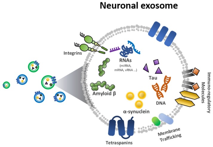
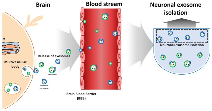
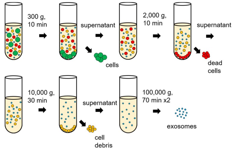
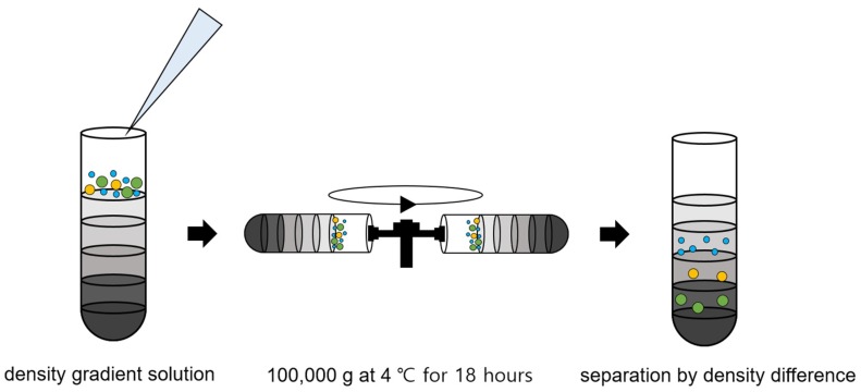
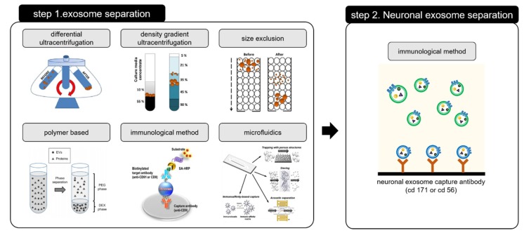
Targeting exosome for liquid biopsy has gained significant attention for its diagnostic and therapeutic potential. For detecting neuronal disease diagnosis such as Alzheimer's disease (AD), the main technique for identifying AD still relies on positron-emission tomography (PET) imaging to detect the presence of amyloid-β (Aβ). While the detection of Aβ in cerebrospinal fluid has also been suggested as a marker for AD, the lack of quantitative measurements has compromised existing assays. In cerebrospinal fluid, in addition to Aβ, T-Tau, and P-Tau, alpha-synuclein has been considered a biomarker of neurodegeneration. This review suggests that and explains how the exosome can be used as a neuronal diagnostic component. To this end, we summarize current progress in exosome preparation/isolation and quantification techniques and comment on the outlooks for neuronal exosome-based diagnostic techniques.

Keywords: Alzheimer’s disease; Parkinson’s disease; diagnostics; exosome.


Conflict of interest statement

The authors declare no conflict of interest.

Figures

Figure 1
Figure 1
Biological elements in a neuronal exosome, including amyloid-β (Aβ), tau and α-synuclein.
Figure 2
Figure 2
Schemes of neuronal exosomes isolation/detection from blood.
Figure 3
Figure 3
Differential ultracentrifugation process for exosome isolations.
Figure 4
Figure 4
Density gradient ultracentrifugation process for exosome isolation.
Figure 5
Figure 5
Size exclusion methods. (a) filtration [42], (b) size exclusion chromatography [46], and (c) combination of filtration and ultracentrifugation [45]. Reprinted with permission from [42,45,46].
Figure 5
Figure 5
Size exclusion methods. (a) filtration [42], (b) size exclusion chromatography [46], and (c) combination of filtration and ultracentrifugation [45]. Reprinted with permission from [42,45,46].
Figure 6
Figure 6
Polymer based exosome isolation of (a) polyethylene glycol (PEG) [52] and (b) aqueous two-phase system (ATPS) [37]. Reprinted with permission from [37,52].
Figure 7
Figure 7
Immunological exosome isolation techniques using enzyme-linked immunosorbent assay (ELISA) [54]. Reprinted with permission from [54].
Figure 8
Figure 8
Exosome isolation using microfluidic platform using (a) microstructure [57], (b) acoustic wave [58], (c) immunological separation [60], and (d) size based separation [62]. Reprinted with permission from [57,58,60,62].
Figure 9
Figure 9
Exosome quantification methods with (a) immunoaffinity capture (IAC) [59], (b) asymmetrical flow field-flow fractionation (AF4) [63], (c) nanoparticle tracking analysis (NTA) [64] (d) dynamic light scattering (DLS) [65], and (e) surface plasmon resonance (SPR) [66]. Reprinted with permission from [59,63,64,66].
Figure 10
Figure 10
Biomarker detection in exosome for Alzheimer’s disease (AD) and Parkinson’s disease (PD). (a) Impedimetric Aβ peptide detection [76]. (b) Carbon nanotube-based field effect transistor (FET) for Aβ detection [78]. (c) Impediemtric microelectrodes for Aβ detection in mouse plasma [11]. (d,e) Electrochemical detection of α-synuclein [79,80]. (f) electrolyter-gated FET for α-synuclein detection [81]. Reprinted with permission from [11,76,78,79,80,81].
Figure 11
Figure 11
Two-step process requiring for isolating the neuronal exosome. Step 1 for exosome separation [46,49,102] and Step 2 for neuronal exosome separation. Reprinted with permission from [46,49,102].

References

    1. Mattson M.P., Pedersen W.A., Duan W., Culmsee C., Camandola S. Cellular and Molecular Mechanisms Underlying Perturbed Energy Metabolism and Neuronal Degeneration in Alzheimer’s and Parkinson’s Diseases. Ann. N. Y. Acad. Sci. 1999;893:154–175. doi: 10.1111/j.1749-6632.1999.tb07824.x. - DOI - PubMed
    1. Taylor C.A., Greenlund S.F., McGuire L.C., Lu H., Croft J.B. Deaths from Alzheimer’s Disease—United States, 1999–2014. MMWR. Morb. Mortal. Wkly. Rep. 2017;66:521–526. doi: 10.15585/mmwr.mm6620a1. - DOI - PMC - PubMed
    1. Alzheimer’s Association 2017 Alzheimer’s disease facts and figures. Alzheimer’s Dement. 2017;13:325–373. doi: 10.1016/j.jalz.2017.02.001. - DOI
    1. Liu S., Liu S., Cai W., Pujol S., Kikinis R., Feng D. Early diagnosis of Alzheimer’s disease with deep learning; Proceedings of the 2014 IEEE 11th International Symposium on Biomedical Imaging (ISBI); Beijing, China. 29 April–2 May 2014; pp. 1015–1018.
    1. Small G.W. Early diagnosis of Alzheimer’s disease: Update on combining genetic and brain-imaging measures. Dialogues Clin. Neurosci. 2000;2:241–246. - PMC - PubMed

LinkOut - more resources