Review
. 2018 Nov 30;25(1):87.
doi: 10.1186/s12929-018-0487-4.

Mitophagy, a potential therapeutic target for stroke

Affiliations
Review

Mitophagy, a potential therapeutic target for stroke

Ruiqiao Guan et al. J Biomed Sci. .

Abstract

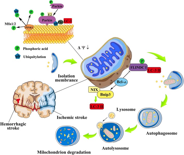
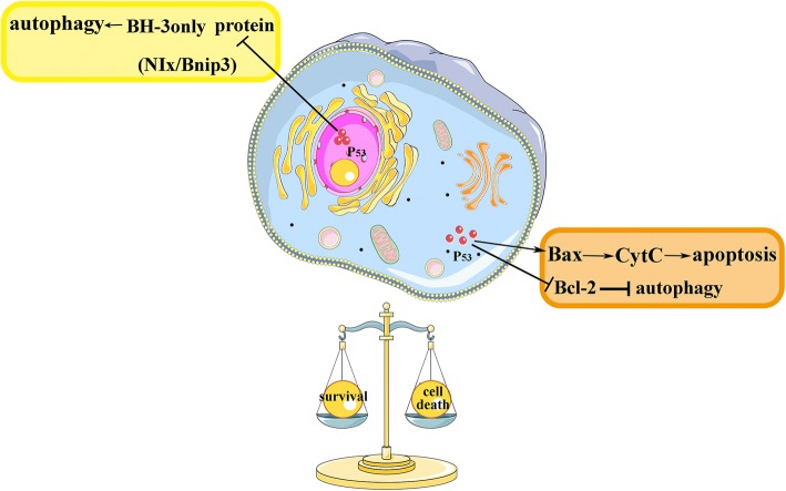
Mitochondria autophagy, termed as mitophagy, is a mechanism of specific autophagic elimination of mitochondria. Mitophagy controls the quality and the number of mitochondria, eliminating dysfunctional or excessive mitochondria that can generate reactive oxygen species (ROS) and cause cell death. Mitochondria are centrally implicated in neuron and tissue injury after stroke, due to the function of supplying adenosine triphosphate (ATP) to the tissue, regulating oxidative metabolism during the pathologic process, and contribution to apoptotic cell death after stroke. As a catabolic mechanism, mitophagy links numbers of a complex network of mitochondria, and affects mitochondrial dynamic process, fusion and fission, reducing mitochondrial production of ROS, mediated by the mitochondrial permeability transition pore (MPTP). The precise nature of mitophagy's involvement in stroke, and its underlying molecular mechanisms, have yet to be fully clarified. This review aims to provide a comprehensive overview of the integration of mitochondria with mitophagy, also to introduce and discuss recent advances in the understanding of the potential role, and possible signaling pathway, of mitophagy in the pathological processes of both hemorrhagic and ischemic stroke. The author also provides evidence to explain the dual role of mitophagy in stroke.

Keywords: Mitochondria; Mitochondria autophagy; Stroke.


Conflict of interest statement

Authors’ information

WZ is Vice chairman of China Institute of traditional Chinese medicine,encephalopathy branch; Vice chairman of World Federation of traditional Chinese Medicine, Mental disorder branch; Vice chairman of Heilongjiang province Association of Acupuncture and Moxibustion; Vice chairman of Association of the Integration of Traditional and Western Medicine and: Association of Traditional Chinese, neurology branch in Heilongjiang province; Expert reviewer of National Awards for science and technology .

Ethics approval and consent to participate

Not applicable.

Consent for publication

Not applicable.

Competing interests

The authors declare that they have no competing interests.

Publisher’s Note

Springer Nature remains neutral with regard to jurisdictional claims in published maps and institutional affiliations.

Figures

Fig. 1
Fig. 1
The molecular mechanism behind mitophagy after stroke. PINK, Parkin, NIX, BNIP3, and the newly-found FUNDC1 are mitophagy receptors in mammalian cells. Mitophagy regulated by PINK1-Parkin-mediated pathway is a multi-step process. Briefly, PINK1 is accumulated on the outer membrane of dysfunctional mitochondria, subsequently to recruit parkin by activating parkin’s cytosolic E3 ubiquitin ligase, then proteins on the outer mitochondrial membrane such as VDAC1 and Mfn1/2 are ubiquitinated by parkin to induce mitophagy. Adapter proteins including p62 are accumulated in the outer mitochondrial membrane after the ubiquitination, leading to ubiquitylated cargo recruited into autophagosomes by binding to LC3. BNIP3 and NIX are related multi-functional outer mitochondrial membrane proteins. Bnip3 and NIX activated mitophagy by binding to Bcl-2 family proteins (including BCL2 and BCL-xL), and also by repressing mTOR or regulating the production of ROS. The molecular mechanism of FUNDC1-mediated mitophagy has not been reported in the pathologies of stroke so far. FUNDC1 phosphorylated at serine 17 under hypoxia stress, thereby to interacting with LC3 and promoting mitophagy. Bcl-xL can inhibit LC-3II conversion, thereby suppressing FUNDC1-mediated mitophagy. In response to the stress of stroke (both of ischemic and hemorrhagic stroke), mitophagy is activated as a stress adaptation to removing dysfunctional mitochondria. Elongated mitochondria were divided into pieces, of which the process is preceded by mitochondrial fission, then autophagosomes, double-membrane vesicles are formed, and sequester targeted cell constituents and mitochondria. The mature autophagosomes then fuse with a lysosome to form the autophagolysosomes, where the mitochondria are subsequently degraded
Fig. 2
Fig. 2
P53 and the dual role of mitophagy. P53 has overlapping alterations with Bcl-2 family protein. P53-involved intrinsic pathway of apoptosis, target Bcl-2 proteins and genes such as Bax, and subsequently induce mitochondrial membrane disruption and cytochrome c release. In autophagy regulation, nuclear P53 would induce autophagy by taking transcriptional effects, like transactivating BH3-only proteins in response to stress; however cytoplasmic P53 might conduct as a major repressor of autophagy by re-localizing to mitochondria, and binding to anti-apoptotic Bcl-2 family members (or activating the proapoptotic Bcl-2 family proteins). Thus, P53 may act to balance autophagy (mitophagy) and apoptosis in regulating cell survival and death in response to stress like stroke injury

References

    1. Jimenez Caballero PE. Spontaneous intracerebral hemorrhage: current perspectives. Medicina clinica. 2014;142(1):23–24. doi: 10.1016/j.medcli.2013.04.032. - DOI - PubMed
    1. Xi G, Keep RF, Hoff JT. Mechanisms of brain injury after intracerebral haemorrhage. The Lancet Neurology. 2006;5(1):53–63. doi: 10.1016/S1474-4422(05)70283-0. - DOI - PubMed
    1. Cervera A, Amaro S, Chamorro A. Oral anticoagulant-associated intracerebral hemorrhage. J Neurol. 2012;259(2):212–224. doi: 10.1007/s00415-011-6153-3. - DOI - PubMed
    1. Kuramatsu JB, Huttner HB, Schwab S. Advances in the management of intracerebral hemorrhage. J Neural Transm. 2013;120(Suppl 1):S35–S41. doi: 10.1007/s00702-013-1040-y. - DOI - PubMed
    1. Berkowitz AL, Mittal MK, McLane HC, Shen GC, Muralidharan R, Lyons JL, Shinohara RT, Shuaib A, Mateen FJ. Worldwide reported use of IV tissue plasminogen activator for acute ischemic stroke. Int J Stroke. 2014;9(3):349–355. doi: 10.1111/ijs.12205. - DOI - PubMed