. 2018 Dec 1;16(1):99.
doi: 10.1186/s12951-018-0424-4.

Specific cancer stem cell-therapy by albumin nanoparticles functionalized with CD44-mediated targeting

Affiliations

Specific cancer stem cell-therapy by albumin nanoparticles functionalized with CD44-mediated targeting

Yuanyuan Li et al. J Nanobiotechnology. .

Abstract

Background: Cancer stem cells (CSCs) are highly proliferative and tumorigenic, which contributes to chemotherapy resistance and tumor occurrence. CSCs specific therapy may achieve excellent therapeutic effects, especially to the drug-resistant tumors.

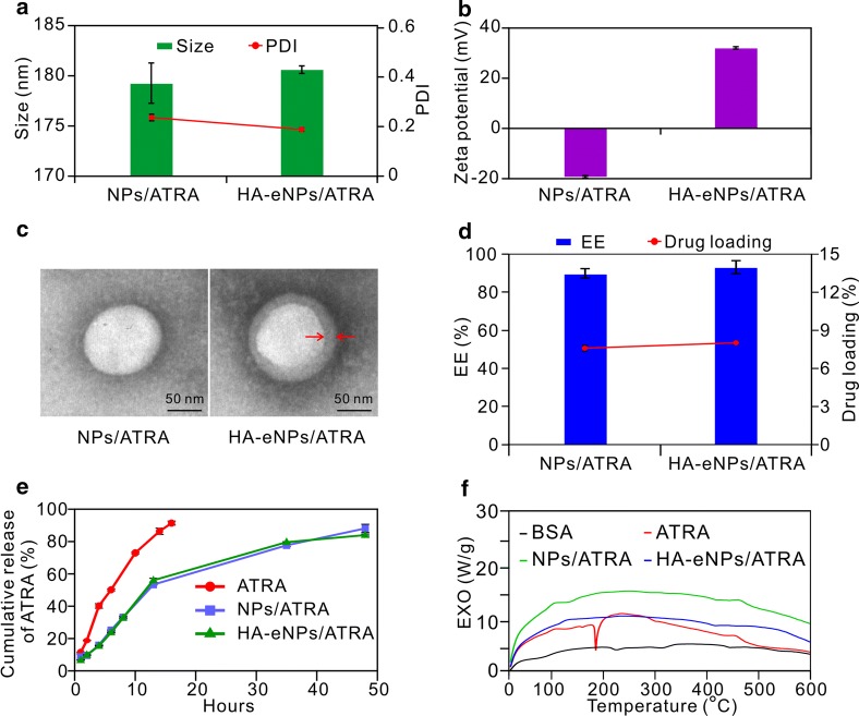
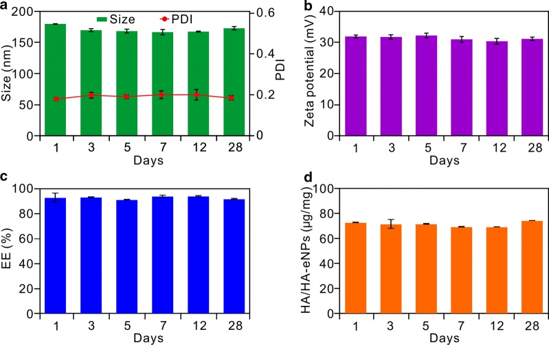
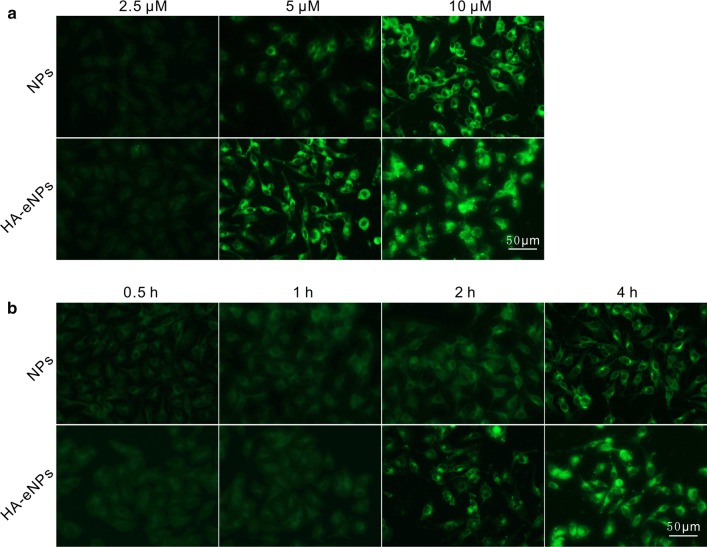
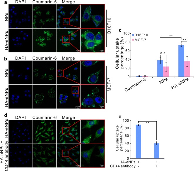
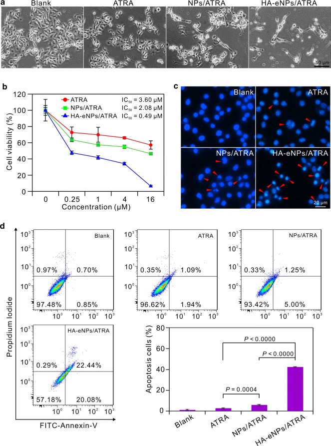
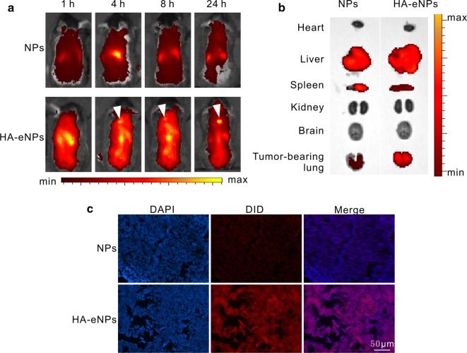
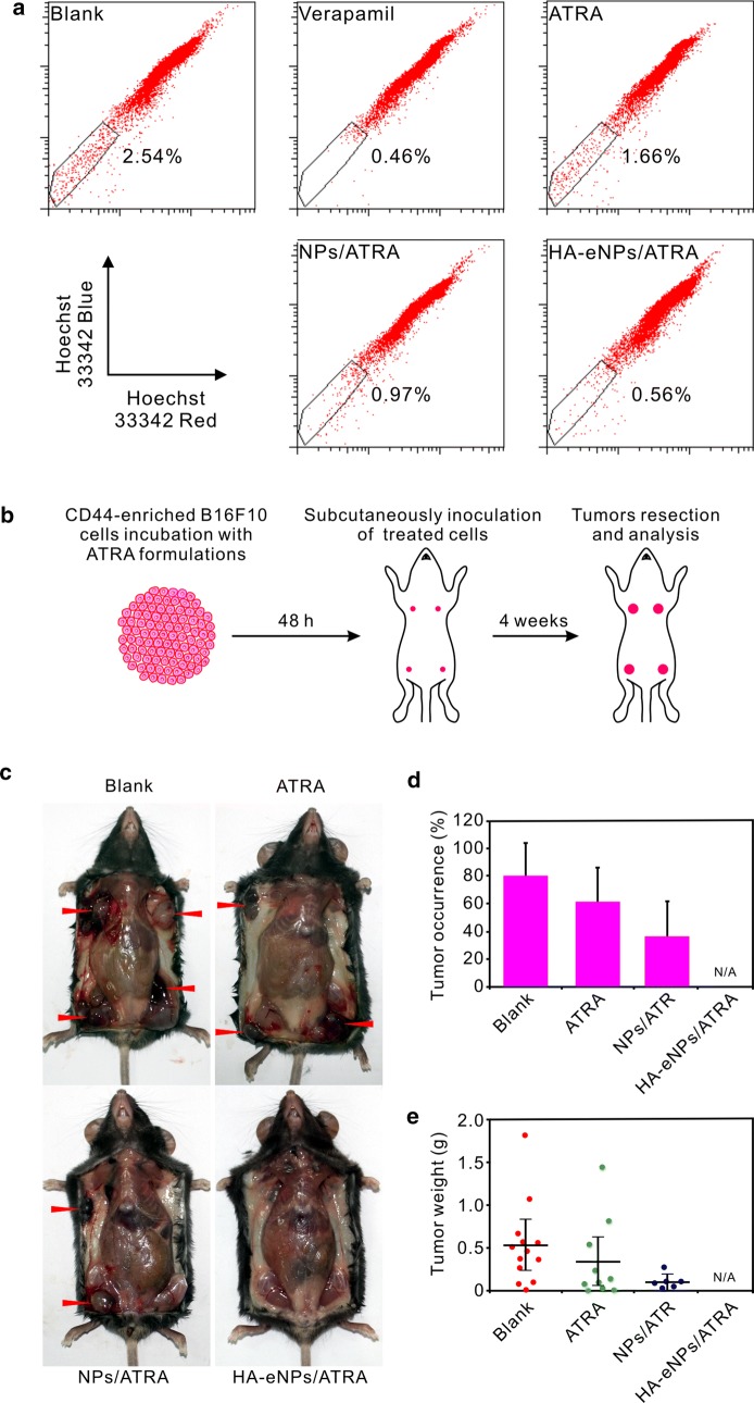
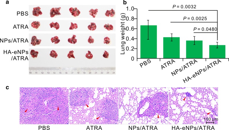
Results: In this study, we developed a kind of targeting nanoparticle system based on cationic albumin functionalized with hyaluronic acid (HA) to target the CD44 overexpressed CSCs. All-trans-retinoic acid (ATRA) was encapsulated in the nanoparticles with ultrahigh encapsulation efficiency (EE%) of 93% and loading content of 8.37%. TEM analysis showed the nanoparticles were spherical, uniform-sized and surrounded by a coating layer consists of HA. Four weeks of continuously measurements of size, PDI and EE% revealed the high stability of nanoparticles. Thanks to HA conjugation on the surface, the resultant nanoparticles (HA-eNPs) demonstrated high affinity and specific binding to CD44-enriched B16F10 cells. In vivo imaging revealed that HA-eNPs can targeted accumulate in tumor-bearing lung of mouse. The cytotoxicity tests illustrated that ATRA-laden HA-eNPs possessed better killing ability to B16F10 cells than free drug or normal nanoparticles in the same dose, indicating its good targeting property. Moreover, HA-eNPs/ATRA treatment decreased side population of B16F10 cells significantly in vitro. Finally, tumor growth was significantly inhibited by HA-eNPs/ATRA in lung metastasis tumor mice.

Conclusions: These results demonstrate that the HA functionalized albumin nanoparticles is an efficient system for targeted delivery of antitumor drugs to eliminate the CSCs.

Keywords: All-trans-retinoic acid; CD44; Cancer stem cells; Cationic albumin; Hyaluronic acid.


Figures

Fig. 1
Fig. 1
In vitro characteristics of NP formulations. a, b Diameter and zeta potential measurements of NP formulations. c Different nanoparticles visualized by transmission electron microscopy (TEM), scale bar: 50 nm (arrows indicate the coated HA layer). d The encapsulation efficiency and drug loading of NP formulations. Results are shown as the means ± SD (n = 3). e Release profiles of ATRA from NP formulations in PBS with 10% (v/v) ethanol. Results are shown as the means ± SD (n = 3). f Differential scanning calorimetry (DSC) thermograms of ATRA formulations
Fig. 2
Fig. 2
Stabilities evaluation of ATRA loaded HA-eNPs. Continually determinations of a the diameter, b the zeta potential, c the encapsulation efficiency, and d HA density of HA-eNPs within 4 weeks, respectively. Results are shown as the means ± SD (n = 3)
Fig. 3
Fig. 3
Fluorescence analysis of uptake of HA-eNPs/ATRA in B16F10 cells. a Fluorescence images of uptake of coumarin-6-labeled NPs at different ATRA concentrations (4 h). b Fluorescence images of uptake of coumarin-6-labeled NPs at different time points (5 μM ATRA). Fluorescence signals were observed by fluorescence microscopy. Scale bar represents 50 μm
Fig. 4
Fig. 4
In vitro evaluation of detecting the targeting of HA-eNPs to CD44. a, b Coumarin-6 loaded HA-eNPs were incubated with CD44-enriched B16F10 cells or CD44-low expressing MCF-7 cells, respectively. Fluorescence signals were observed by confocal microscopy. c Flow cytometry analysis showing the cellular uptake percentage of the B16F10 and MCF-7 cells after incubation with free coumarin-6, coumarin-6 loaded NPs, or HA-eNPs. Data are shown as the means ± SD, **P < 0.01, ns not significant (n = 3). d, e B16F10 cells were preincubated with anti-CD44 antibody and followed by incubating with HA-eNPs for 4 h. Fluorescence signals were observed by confocal microscopy and flow cytometry. Data are shown as the means ± SD, **P < 0.01 (n = 3)
Fig. 5
Fig. 5
In vitro antitumor effects of NP formulations on the B16F10 cells. a Morphological changes of B16F10 cells after treatment with various ATRA formulations. Scale bar represents 50 μm. b MTT viability assay for B16F10 cells after treatment with various ATRA formulations. Results are shown as the means ± SD (n = 3). c Apoptosis of B16F10 cells induced by various ATRA formulations (5 μM) (arrows indicate cell apoptosis). Scale bar represents 20 μm. d Flow cytometry analysis of B16F10 cell apoptosis induced by ATRA formulations. Results are shown as the means ± SD (n = 3)
Fig. 6
Fig. 6
Fluorescence imaging of ATRA-loaded NP formulations in mice. a Time-dependent intensity images of fluorescence distribution in mice (arrows indicate accumulation of HA-eNPs in tumor-bearing lungs). b Fluorescence images of major organs at 8 h post-injection of NP formulations in mice. c Fluorescence microscopy images of tumor-bearing lung tissue section. Scale bar represents 50 μm
Fig. 7
Fig. 7
HA-eNPs/ATRA decreased the tumorigenicity of CD44-enriched B16F10 cells. a Side population (SP) changes in B16F10 cells after treatment with various ATRA formulations. b A diagram of the tumorigenicity assessment. In the experiment, four flanks of each mouse were injected with the same treated or untreated B16F10 cells. c Representative photo of tumors 24 days after subcutaneous implantations of ATRA formulations treated or untreated B16F10 cells (arrows indicate tumor nodules). d, e Tumor occurrences and weights measured 24 day after implantation. Results are shown as the means ± SD (n = 16)
Fig. 8
Fig. 8
HA-eNPs/ATRA enhanced the inhibition of tumor growth in the in situ lung metastasis tumor-bearing mice. a Images of the B16F10-bearing lungs on day 24 after five consecutive i.v. injections of NP formulations (n = 5). b Antitumor effects of various treatments evaluated according to B16F10-bearing lung weight (n = 5). c Histological staining of the B16F10-bearing lungs after various treatments (arrows indicate tumor nodules, n = 5)
Fig. 9
Fig. 9
Safety evaluation of ATRA-loaded NP formulations. Microscopic images of hematoxylin and eosin (H&E) staining of organs (liver, spleen, lung, kidney, heart and brain) from mice treated with ATRA-loaded NP formulations. No abnormality was observed. Scale bar represents 50 μm

References

    1. Lortettieulent J, Soerjomataram I, Ferlay J, Rutherford M, Weiderpass E, Bray F. International trends in lung cancer incidence by histological subtype: adenocarcinoma stabilizing in men but still increasing in women. Lung Cancer. 2014;84:13–22. doi: 10.1016/j.lungcan.2014.01.009. - DOI - PubMed
    1. Siegel RL, Miller KD, Jemal A. Cancer statistics, 2018. Cancer J Clin. 2018;68:7–30. doi: 10.3322/caac.21442. - DOI - PubMed
    1. Jordan CT, Guzman ML, Noble M. Cancer stem cells. N Engl J Med. 2006;355:1253–1261. doi: 10.1056/NEJMra061808. - DOI - PubMed
    1. Nandy SB, Lakshmanaswamy R. Cancer stem cells and metastasis. Prog Mol Biol Transl Sci. 2017;151:137–176. doi: 10.1016/bs.pmbts.2017.07.007. - DOI - PubMed
    1. Reya T, Morrison SJ, Clarke MF, Weissman IL. Stem cells, cancer, and cancer stem cells. Nature. 2001;414:105. doi: 10.1038/35102167. - DOI - PubMed

LinkOut - more resources