. 2018 Dec 18;115(12):2336-2347.
doi: 10.1016/j.bpj.2018.10.031. Epub 2018 Nov 10.

Cooperative Induction of Ordered Peptide and Fatty Acid Aggregates

Affiliations

Cooperative Induction of Ordered Peptide and Fatty Acid Aggregates

Radoslaw Bomba et al. Biophys J. .

Abstract

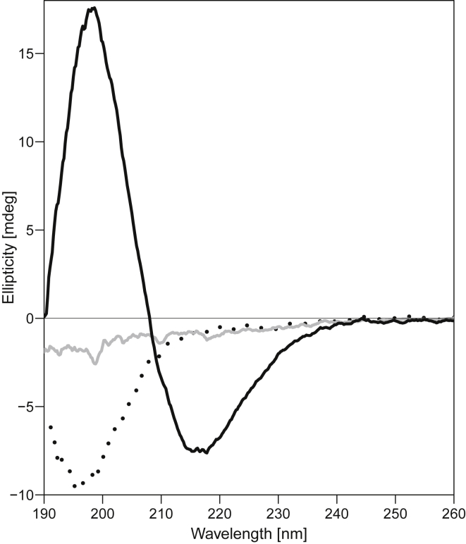
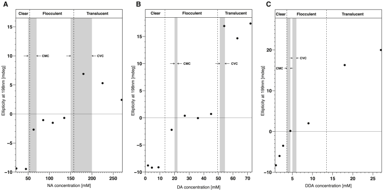
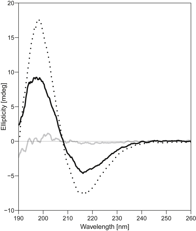
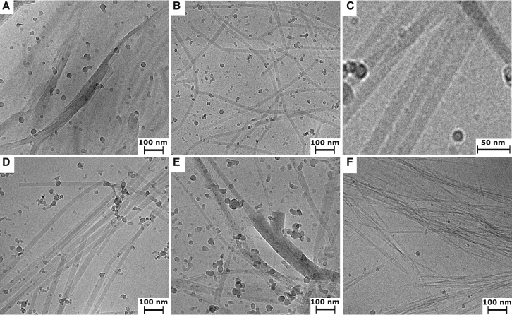
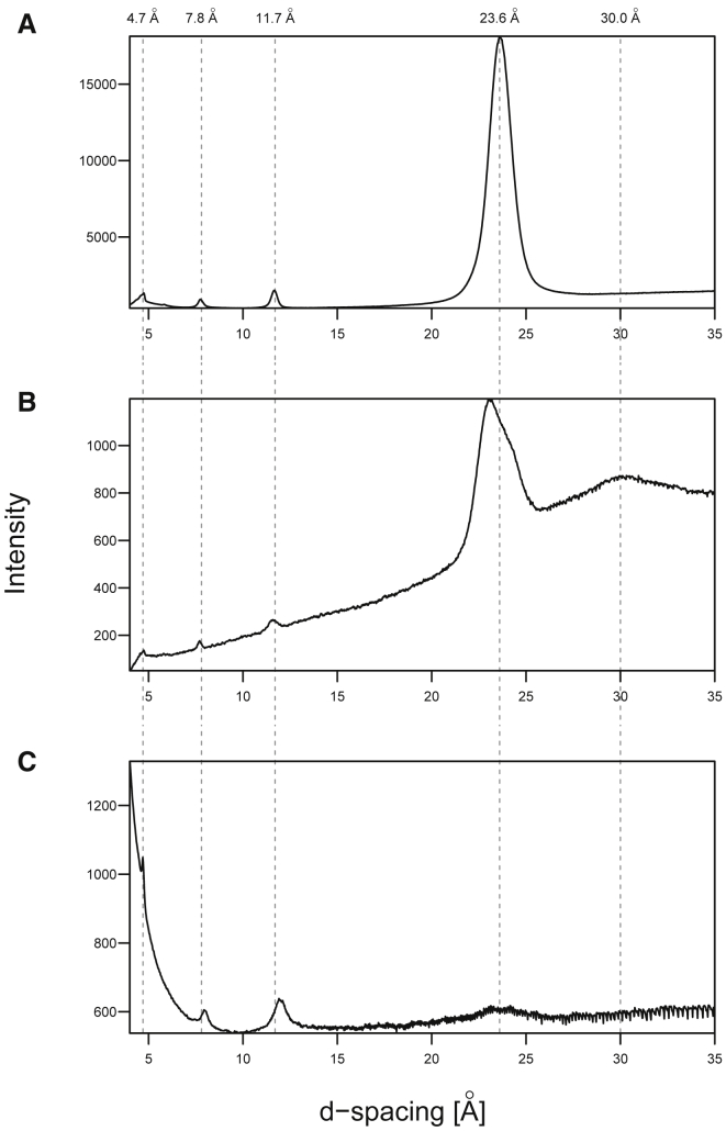
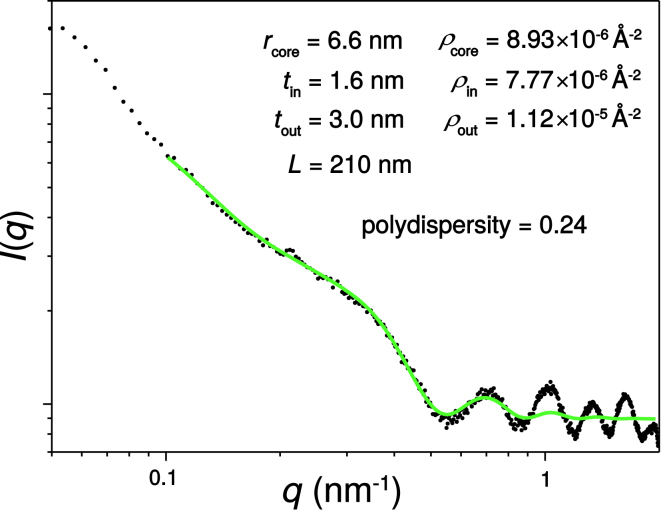
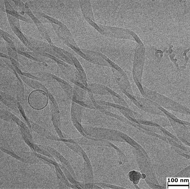
Interactions between biological membranes and disease-associated amyloids are well documented, and their prevalence suggests that an inherent affinity exists between these molecular assemblies. Our interest in the molecular origins of life have led us to investigate the nature of such interactions in the context of their molecular predecessors (i.e., vesicle-forming amphiphiles and small peptides). Under certain conditions, amyloidogenic peptides or fatty acids are each able to form ordered structures on their own; however, we report here on their cooperative assembly into novel, to our knowledge, highly ordered structures. We first examined an amyloidogenic eight-residue peptide, which forms amyloids at pH 11, yet because of its positive electrostatic character remains soluble at a neutral pH. In mixtures with simple fatty acids, this peptide is also able to form novel, to our knowledge, coaggregates at a neutral pH whose structures are sensitive to both the fatty acid concentration and identity. Below the critical vesicle concentration, the mixtures of fatty acid and peptide yield a flocculent precipitate with an underlying β-structure. Above the critical vesicle concentration, the mixtures yield a translucent precipitate that consists of tube-like structures. Small-angle x-ray scattering and fiber diffraction data were used to model their structures as hollow-core two-shell cylinders in which the inner shell is a bilayer of fatty acid and the outer shell alternates between amyloid and bilayers of fatty acid. The further analysis of decanoic acid with a panel of 13 other basic amyloidogenic peptides confirmed the general nature of the observed interactions. The cooperativity within this heterogeneous system is attributed to the structurally repetitive natures of the fatty acid bilayer and the cross-β-sheet motif, providing compatible scaffolds for attractive electrostatic interactions. We show these interactions to be mutually beneficial, expanding the phase space of both peptides and fatty acids while providing a simple yet robust physical connection between two distinct entities relevant for life.


Figures

Figure 1
Figure 1
The concentration dependence of the CD spectra of DA solutions with (OV)4. The CD spectra of three solutions of 575 μM (OV)4 with either 1.8 mM DA (dotted line), 18 mM DA (solid gray line), or 72 mM DA (solid black line) are shown. At 1.8 mM DA, the CD spectrum is random coil like; at 18 mM DA, it is nearly featureless; and at 72 mM, it is β-sheet like.
Figure 2
Figure 2
Correlation of (OV)4 secondary structure with fatty acid CMC and CVC. The β-strand secondary structure of the peptide in the fatty acid mixtures as monitored by its CD signal at 198 nm is plotted against the fatty acid concentration in solutions of NA (A), DA (B), and DDA (C). Negative values are associated with a random coil-like structure of the peptide and positive values with a β-sheet secondary structure. The dashed lines roughly demarcate the boundaries between the visible changes in the mixtures from clear solution to flocculent precipitate to translucent solution with increasing concentration of fatty acid. The gray bands represent the ranges of CMC and CVC values for the fatty acids as measured independently in the conditions used in this study (see Figs. S3–S6) and indicate that the transitions of peptide/fatty acids mixtures occur at very similar concentration ranges as do the phase transitions of the pure fatty acids themselves. The deviation of the DDA mixtures from this trend can be explained by the fact that its concentration is low enough that the binding of DDA to the 575 μM (OV)4 consumes a significant amount of the fatty acid, effectively shifting the flocculent-translucent boundary.
Figure 3
Figure 3
Fatty alcohol lowers both CVC and the concentration at which translucent precipitate is formed. Depicted are three CD spectra for 575 μM solutions of (OV)4 with either 72 mM DA (dotted line), 36 mM DA (solid gray line), or a 10:1 DA/DOH mixture with 34.65 mM total amphiphiles (solid black line). The β-sheet-like spectrum absent in the 36 mM DA solution due to the formation of the flocculent precipitate is present in the lower concentration DA solution with a 1:10 molar ratio of fatty alcohol. This amount of DOH decreases the CVC of the system from ∼55 to ∼20 mM (Fig. S10).
Figure 4
Figure 4
Morphology of fatty-acid-(OV)4 precipitates compared to pure (OV)4 amyloids. Cryo-EM micrographs of the flocculent precipitate (A) and translucent precipitate (B) formed in mixtures of DA and (OV)4. The flocculent precipitate is usually too thick for observing detailed morphological features, but in a few instances like the one depicted in (A), fibrillar structures are visible. The translucent precipitate is characterized by straight fiber-like structures that have a uniform width of ∼25 nm and lengths of up to several microns. In many of the tube-like structures, there is a visible striation, indicating that they may be composed of an alignment of smaller fibrils (C). The translucent precipitates of (OV)4 mixtures with NA (D) and DDA (E) also reveal fiber-like structures; however, they present a distribution of widths. The NA samples have a narrow distribution, whereas the DDA/(OV)4 mixtures have structures with a much larger variation in width. The amyloid fibrils of (OV)4 that form at pH 11 (F) are characterized by many straight fibers that form bundles of varying numbers of protofilaments.
Figure 5
Figure 5
X-ray diffraction of fatty acid-(OV)4 precipitates compared to pure (OV)4 amyloids. The x-ray diffraction images of flocculent (A) and translucent (B) precipitates from DA-(OV)4 mixtures at 18 mM DA and 63 mM DA, respectively, are shown. (C) The diffraction of (OV)4 amyloids formed at pH 11 in the absence of fatty acids is shown. All of the various aggregates have a reflection at 4.7 Å corresponding to the spacing between strands in a β-sheet. The other similarities include reflections near 7.8 and 11.7 Å. The DA/(OV)4 precipitates also have an intense reflection around 24 Å that is mostly absent in the pH 11 amyloid fibrils that lack DA. Unique to the translucent precipitate is a broad low-resolution reflection at 30 Å. The diffraction patterns were radially integrated with the FIT2D software tο yield a plot of the scattered intensity as a function of d-spacing.
Figure 6
Figure 6
A one-dimensional SAXS intensity profile for the DA-(OV)4 translucent precipitate. The intensity of the scattering (I) as a function of the scattering wave vector (q) is plotted with the fit to the hollow-core two-shell cylinder form factor P(q) (see Supporting Materials and Methods) in green. The fitted parameters for this form factor are listed in the figure. To see this figure in color, go online.
Figure 7
Figure 7
A cryo-EM image of DFRFRF-NH2 peptide mixture with 72 mM DA at pH 7.8. The twisted ribbons resulting in the shape of helices are formed in these conditions. The widths of the ribbons as well as the tightness of helices varies between each assembly. A vesicle of decanoic acid (DA) is also visible in the image.

Similar articles

Cited by

References

    1. Orgel L.E. Evolution of the genetic apparatus. J. Mol. Biol. 1968;38:381–393. - PubMed
    1. Wächtershäuser G. Before enzymes and templates: theory of surface metabolism. Microbiol. Rev. 1988;52:452–484. - PMC - PubMed
    1. Segré D., Ben-Eli D., Lancet D. The lipid world. Orig. Life Evol. Biosph. 2001;31:119–145. - PubMed
    1. Gilbert W. Origin of life: the RNA world. Nature. 1986;319:618.
    1. Oba T., Fukushima J., Ikehara K. Catalytic activities of [GADV]-peptides. Formation and establishment of [GADV]-protein world for the emergence of life. Orig. Life Evol. Biosph. 2005;35:447–460. - PubMed

LinkOut - more resources