Review
. 2019 Feb:43:23-36.
doi: 10.1016/j.smrv.2018.09.006. Epub 2018 Nov 8.

Neurobiological and immunogenetic aspects of narcolepsy: Implications for pharmacotherapy

Affiliations
Review

Neurobiological and immunogenetic aspects of narcolepsy: Implications for pharmacotherapy

Steven T Szabo et al. Sleep Med Rev. 2019 Feb.

Abstract

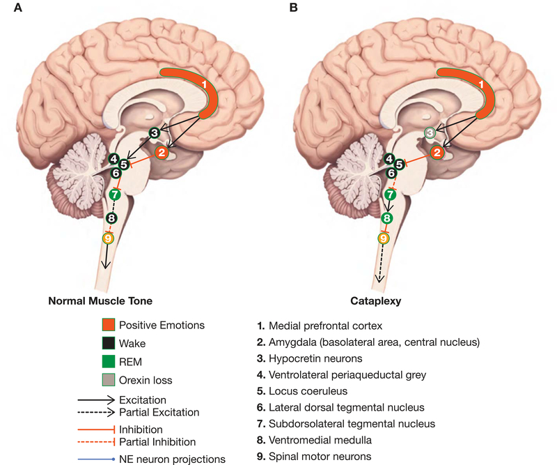
Excessive daytime sleepiness (EDS) and cataplexy are common symptoms of narcolepsy, a sleep disorder associated with the loss of hypocretin/orexin (Hcrt) neurons. Although only a few drugs have received regulatory approval for narcolepsy to date, treatment involves diverse medications that affect multiple biochemical targets and neural circuits. Clinical trials have demonstrated efficacy for the following classes of drugs as narcolepsy treatments: alerting medications (amphetamine, methylphenidate, modafinil/armodafinil, solriamfetol [JZP-110]), antidepressants (tricyclic antidepressants, selective serotonin reuptake inhibitors, serotonin-norepinephrine reuptake inhibitors), sodium oxybate, and the H3-receptor inverse agonist/antagonist pitolisant. Enhanced catecholamine availability and regulation of locus coeruleus (LC) norepinephrine (NE) neuron activity is likely central to the therapeutic activity of most of these compounds. LC NE neurons are integral to sleep/wake regulation and muscle tone; reduced excitatory input to the LC due to compromise of Hcrt/orexin neurons (likely due to autoimmune factors) results in LC NE dysregulation and contributes to narcolepsy/cataplexy symptoms. Agents that increase catecholamines and/or LC activity may mitigate EDS and cataplexy by elevating NE regulation of GABAergic inputs from the amygdala. Consequently, novel medications and treatment strategies aimed at preserving and/or modulating Hcrt/orexin-LC circuit integrity are warranted in narcolepsy/cataplexy.

Keywords: Amygdala; Autoimmune; Cataplexy; Excessive daytime sleepiness; Hypocretin; Locus coeruleus; Mechanism of action; Narcolepsy; Norepinephrine; Orexin.


Conflict of interest statement

Conflicts of interest

Dr. Szabo has an investigator-initiated grant from Otsuka Pharmaceuticals and funding support from the National Institutes of Health (NIH) and the Brain Behavior Research Foundation (formerly known as NARSAD). He has served on the advisory board for Jazz Pharmaceuticals and as a consultant/speaker for Neurocrine Biosciences, Teva Pharmaceutical Industries Ltd, and Otsuka/Lundbeck Pharmaceuticals.

Dr. Thorpy has received research/grant support and consultancy fees from Jazz Pharmaceuticals, Avadel Pharmaceuticals, Harmony Biosciences, Balance Therapeutics, and Merck Inc.

Dr. Peever has received funding from the National Science and Engineering Research Council and the Canadian Institutes of Health Research.

Dr. Mayer has received honoraria from the Paul Ehrlich Institute, Germany, has served on the speakers’ bureau for UCB Pharma and Sanofi, and is a board member of the European Narcolepsy Network.

Dr. Kilduff has received funding for narcolepsy-related research from the National Institutes of Health, Jazz Pharmaceuticals and TevaBranded Pharmaceuticals Products R&D, Inc.

Figures

Fig. 1.
Fig. 1.. Circuit mechanisms controlling cataplexy and Rapid Eye Movement (REM) sleep atonia.
(Sagittal section of the human brain adapted from [103]). The subdorsolateral tegmental nucleus (SLD; 7) and ventromedial medulla (VM; 8) constitute the core circuits that generate rapid eye movement (REM) sleep atonia. When (presumably glutamatergic) SLD neurons become active, they stimulate gamma-aminobutyric acid (GABA)/glycine neurons in the VM such that they trigger REM sleep atonia by hyperpolarizing spinal motor neurons (MN; 9). A) In non-narcoleptics, positive emotions do not trigger muscle atonia because descending inhibition from GABAergic neurons in the central nucleus of the amygdala (CeA; 2) to the locus coeruleus (LC), laterodorsal tegmentum/pedunculopontine nuclei (LDT/PPT), and ventrolateral periaqueductal gray (vlPAG) is offset by excitatory hypocretin/orexin inputs (3), which prevent positive emotions from accessing the REM sleep circuits (i.e., SLD => VM) that trigger muscle atonia. B) In narcolepsy, there is decreased excitatory Hct input (3) to LC/LDT/PPT and vlPAG. Positive emotions activate cortical areas such as the medial prefrontal cortex (mPFC; 1) which innervate neurons of the basolateral area (BLA; 2) of the amygdala that project to GABA neurons in the CeA. GABAergic CeA neurons then inhibit the LC, LDT/PPT, and vlPAG, which in turn disinhibit the SLD, thereby allowing it to activate the VM to produce MN inhibition and hence muscle atonia/weakness during cataplexy. Abbreviations: BLA, Basolateral area; CeA, Central nucleus of the amygdala; GABA, Gamma-aminobutyric acid; LC, Locus coeruleus; LDT/PPT, Laterodorsal tegmentum/pedunculopontine nuclei; MN, Motor neurons; mPFC, Medial prefrontal cortex; NE, Norepinephrine; REM, Rapid eye movement; SLD, Subdorsolateral tegmental nucleus; vIPAG, Ventrolateral periaqueductal gray; VM: Ventromedial medulla
Fig. 2.
Fig. 2.
A) NE neuron projections (blue) widely innervate the human brain (adapted from [103]). LC NE neurons send ascending projections to the cortex that facilitate wakefulness and descending projections to the spinal cord that are important in maintaining muscle tone. LC NE neurons receive a prominent excitatory projection from Hcrt/orexin neurons in the tuberal hypothalamus. The loss of excitatory drive onto LC NE neurons due to Hcrt/orexin neuron compromise in narcolepsy can lead to EDS (i.e., lack of NE tone to the forebrain). Dysregulated LC NE neurons may also permit reduced muscle tone in response to GABAergic input from the amygdala, resulting in cataplexy in response to emotional stimuli. B) A schematic brainstem circuit (adapted from [59]) depicting 5-HT neurons from the dorsal raphe, as well as glutamate and GABA projections to the LC, which are modulators of NE activity. 5-HT exerts a tonic inhibitory tone on LC NE neuron activity and LC NE neurons provide an excitatory tone to 5-HT neurons (not illustrated). Glutamate and GABA in the LC impacts NE neuron firing directly and by modulating 5-HT input. Medications used in the treatment of narcolepsy increase NE release and/or availability with changes onto LC NE neuron activity. a) SSRIs: Desensitization of 5-HT autoreceptors (5-HT1A and 5-HT1D) during prolonged SSRI treatment leads to increased 5-HT transmission. 1) Desensitization of 5-HT1A heteroceptors on glutamate neuron projections to the LC and increased activation of excitatory amino acid (EAA) receptors on 5-HT terminals (green circle) contributes to 2) increased 5-HT activation of 5-HT2A receptors on GABAergic projections (green bar). 3) SSRIs reduce NE neuron stimulation and activity by increasing GABAA receptor activation in the LC (green bar) and 4) increased 5-HT3 receptor activation (likely on NE terminals) can enhance NE in postsynaptic structures. b) SNRIs/TCAs: In addition to effects as outlined in (a), SNRIs and TCAs also produce an increase in NE availability due to NE reuptake transporter blockade. This results in increased activation of α2-autoreceptors (green pill on NE neurons) and reduced activity and stimulation of LC activity. c) Amphetamines / Sympathomimetics / JZP-110 (solriamfetol): Alerting medications increase the release and/or reuptake of NE and DA (depending on the medication) and produce similar effects on LC NE neuron activity as SNRIs and TCAs. d) GHB/SXB: Activation of GABAB receptors on NE neurons reduces LC activity and discontinuation produces an increase in LC NE neuron activity. Given the short half-life (30 min to 1 hour) of SXB, the nighttime sedative effects wane, and increased LC NE neuron stimulation and activity ensues during the day; hence, its effectiveness in treating both EDS and cataplexy in narcolepsy. e) Pitolisant: Modulation of H3 autoreceptors (inverse agonist/antagonist) on histamine neurons in the tuberomammillary nucleus produce increased histamine release onto LC NE neurons. LC NE neurons increase in activity following binding to H1 and H2 receptors on NE neurons, which is effective in treating both EDS and cataplexy in narcolepsy. Abbreviations: α2, Type 2 alpha adrenergic receptor; DA, Dopamine; D2, Type 2 DA receptor; DAT, DA transporter; EAA, Excitatory amino acid; EDS, Excessive daytime sleepiness; GABA, Gamma-amino-butyric acid; GABAA, Type A GABA receptor; GHB, Gamma-hydroxybutyrate; GLU, Glutamate; HA, Histamine; H1, Type 1 HA receptor; H2, Type 2 HA receptor; H3, Type 3 HA receptor; Hcrt, Hypocretin; 5-HT, Serotonin; 5-HT1A, Type 1A 5-HT receptor; LC, Locus coeruleus; NE, Norepinephrine; SNRI, Serotonin norepinephrine reuptake inhibitor; SSRI, Selective serotonin reuptake inhibitor; SXB, Sodium oxybate; TCA, Tricyclic antidepressant.

References

    1. Thorpy MJ, Krieger AC. Delayed diagnosis of narcolepsy: characterization and impact. Sleep Med. 2014; 15: 502–7. - PubMed
    1. Scammell TE. Narcolepsy. N Engl J Med. 2015; 373: 2654–62. - PubMed
    1. Black J, Reaven NL, Funk SE, McGaughey K, Ohayon M, Guilleminault C, et al. The Burden of Narcolepsy Disease (BOND) study: health-care utilization and cost findings. Sleep Med. 2014; 15: 522–9. - PubMed
    1. Black SW, Yamanaka A, Kilduff TS. Challenges in the development of therapeutics for narcolepsy. Prog Neurobiol. 2017; 152: 89–113. - PMC - PubMed
    1. American_Academy_of_Sleep_Medicine. The International Classification of Sleep Disorders – Third Edition (ICSD-3). Darien, IL: American Academy of Sleep Medicine; 2014.

Publication types

MeSH terms

Substances