Review
. 2019 Feb;60(2):242-268.
doi: 10.1194/jlr.R089730. Epub 2018 Nov 30.

The interface between phosphatidylinositol transfer protein function and phosphoinositide signaling in higher eukaryotes

Affiliations
Review

The interface between phosphatidylinositol transfer protein function and phosphoinositide signaling in higher eukaryotes

Aby Grabon et al. J Lipid Res. 2019 Feb.

Abstract

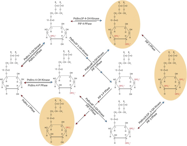
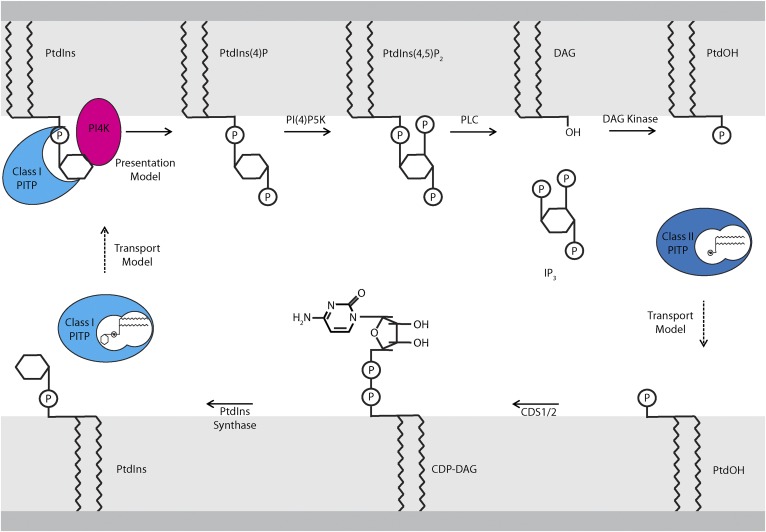
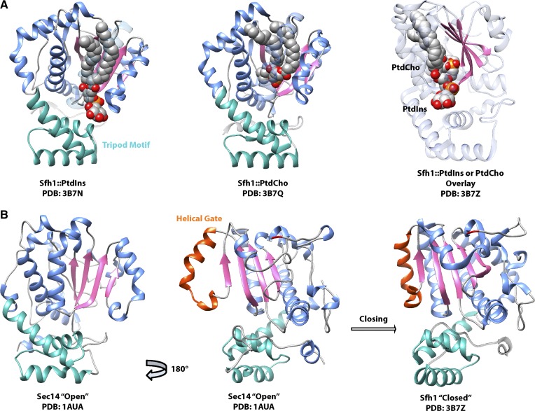
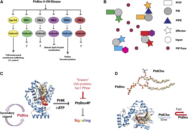
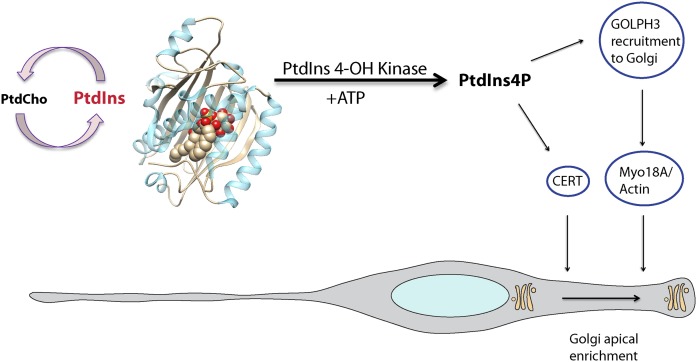
Phosphoinositides are key regulators of a large number of diverse cellular processes that include membrane trafficking, plasma membrane receptor signaling, cell proliferation, and transcription. How a small number of chemically distinct phosphoinositide signals are functionally amplified to exert specific control over such a diverse set of biological outcomes remains incompletely understood. To this end, a novel mechanism is now taking shape, and it involves phosphatidylinositol (PtdIns) transfer proteins (PITPs). The concept that PITPs exert instructive regulation of PtdIns 4-OH kinase activities and thereby channel phosphoinositide production to specific biological outcomes, identifies PITPs as central factors in the diversification of phosphoinositide signaling. There are two evolutionarily distinct families of PITPs: the Sec14-like and the StAR-related lipid transfer domain (START)-like families. Of these two families, the START-like PITPs are the least understood. Herein, we review recent insights into the biochemical, cellular, and physiological function of both PITP families with greater emphasis on the START-like PITPs, and we discuss the underlying mechanisms through which these proteins regulate phosphoinositide signaling and how these actions translate to human health and disease.

Keywords: cell signaling; diseases; lipid and membrane trafficking; lipid signaling; lipids • membranes.


Figures

Fig. 1.
Fig. 1.
Phosphoinositide metabolism. The synthesis and degradation of phosphoinositides by kinases and phosphatases (denoted as “PPase”), respectively, is depicted in schematic form. PLs in tan bubbles exist in higher eukaryotes but not S. cerevisiae.
Fig. 2.
Fig. 2.
Models of PITP function in the PIP cycle. PtdIns is synthesized at the ER by the sequential action of the enzymes, CDS1/2 and PtdIns synthase, which convert PtdOH to CDP-DAG and CDP-DAG to PtdIns, respectively. “Transport models” for describing PITP function postulate that class I PITPs transfer PtdIns from the ER to signaling membranes (i.e., the plasma membrane). “Presentation models” describe PITPs as noncatalytic factors that present PtdIns to PI4K, thereby directly stimulating PtdIns4P synthesis. In the presentation model, PITP utilizes PtdIns at the signaling membrane and does not catalyze PtdIns transport from the ER. PtdIns4P synthesized at the plasma membrane can be converted to PtdIns(4,5)P2, which is then hydrolyzed by agonist-stimulated PLC activity to produce DAG and IP3. DAG is converted to PtdOH by DAG kinase. PtdOH must then be replenished at the ER to restore the PtdIns biosynthetic cycle. Class II PITPs, which bind PtdOH, are proposed to fulfill this role in a transport mechanism.
Fig. 3.
Fig. 3.
The Sec14 structure. Crystal structures of Sec14 and the Sec14-like PITP Sfh1 are shown, with the tripod motif (green) oriented as if toward the membrane. The lipid binding β-sheet floor is rendered in pink, and the helices that line the binding pocket are in blue. A: Crystal structures of the Sec14-like PITP Sfh1 reveal that PtdIns and PtdCho both bind in the lipid binding pocket, with the PtdCho headgroup buried deep within the pocket and the PtdIns headgroup facing outwards toward the protein surface. Overlay of Sfh1 bound to either lipid illustrates that PtdIns and PtdCho acyl chain binding regions overlap, and Sfh1 cannot fully accommodate both lipids at the same time. B: The crystal structure of open lipid-free Sec14 is compared with Sfh1 bound to PtdCho (lipid not shown for clarity). The helical gate (orange) closes around the lipid binding pocket, with a key residue of the G-module (G266) highlighted in red.
Fig. 4.
Fig. 4.
The Sec14 PtdIns presentation mechanism. A: Sec14-like PITPs diversify the biological outcomes of PI4K in cells by specifying unique PtdIns4P pools that promote unique cellular processes. B: Transient complexes that bring together an individual PITP with a PI4K and a set of PtdIns4P effectors, either as individual proteins or in PITP-multidomain arrangements, generate a signaling pixel. The identities of the PITPs in the complex, the specific metabolic input that these sense in the form of the second ligands they bind for priming PtdIns presentation to the PI4K, and the PtdIns4P effectors determine distinct biological outcomes. The pixel boundary is the molecular space of each PITP/PI4K/effector complex. Populating interstitial areas of the membrane with PtdIns4P phosphatases sharpens pixel boundaries and enables PtdIns4P signaling at essentially point resolution. C: Sec14-like PITPs exchange a second ligand for PtdIns, and present PtdIns to PI4K, which generates PtdIns4P used for signaling reactions. The forward reaction is antagonized by PtdIns4P “erasers,” or negative regulators, such as Osh proteins or Sac1 phosphatase. D: PtdIns and PtdCho occupy overlapping positions in the Sec14 lipid-binding pocket. The slow egress of PtdCho from the Sec14 pocket frustrates entry of incoming PtdIns, resulting in an abortive exchange that exposes (presents) the frustrated PtdIns to the PI4K.
Fig. 5.
Fig. 5.
The START PITP family. START PITPs are aligned by primary sequence using VectorNTI (Life Technologies), and the alignment is visualized as a cladogram. Proteins discussed in this review are highlighted in color. Accession numbers for the specific amino acid sequences used are in brackets. Class I PITPs are designated in blue, and class II PITPs in red. PITPs from ancient eukaryotes, including Toxoplasma and Trichomonas do not fall into either category.
Fig. 6.
Fig. 6.
START PITP domain architecture. The class I and class II PITPs discussed in this review are depicted in schematic form with specific domains indicated. Two transcriptional variants each for PITPβ and PITPnc1 are indicated. PITPnm1 is depicted in its full-length wild-type form, as well as the premature truncation found in a subset of the human population [Exome Aggregation Consortium (ExAC) (http://exac.broadinstitute.org/about)].
Fig. 7.
Fig. 7.
START PITP structure. A: The crystal structure of PITPα bound to PtdIns is shown in two orientations as indicated by the arrow. The β-sheet floor of the lipid binding pocket (gray), the regulatory loop (green), helix F (orange), helix A (blue), helix G (purple), and the lipid exchange loop (pink) are highlighted. B: The PtdIns headgroup-binding motif of PITPα is illustrated and the coordinating residues within the lipid binding pocket (inset: Thr59, Lys61, Glu86, Gln90) are highlighted. Residues on helix G that do not bind the headgroup directly but modulate the conformational dynamics that specifically influence PtdIns-binding are also highlighted (cyan: Met240, Glu248). C: The crystal structures of open lipid-free PITPα and PtdCho-bound PITPα are shown to illustrate the conformational dynamics associated with gating of the hydrophobic lipid-binding pocket during the exchange cycle. The lipid exchange loop controls access to the pocket and helix G and helix A fold over the lipid binding floor. Coloring scheme is as in A. D: The crystal structure of PITPα bound to PtdCho is shown with membrane-association regions. Hydrophobic interactions with the membrane are rendered in green and involve residues 135-163 and 259-264, while electrostatic interactions with the membrane involve residues 28-41 and 99-110 and are highlighted in blue. Residues that interact with the membrane only when PtdIns is present in the bilayer are indicated in bright green. The lipid exchange loop is made translucent for the purpose of clarity, and this substructure penetrates the bilayer surface.
Fig. 8.
Fig. 8.
Class I START PITP function in apical loading of the Golgi system in neural stem cells. Class I START PITPs exchange PtdCho and PtdIns, thereby stimulating PI4K activity on late Golgi membranes. The PtdIns4P pool recruits GOLPH3 and CERT to Golgi membranes with GOLPH3 subsequently engaging the apically directed actin machinery via the nonconventional myosin, Myo18A. This interaction promotes loading of the Golgi system to the neural stem cell apical process, thereby setting up an asymmetry critical for neural establishment/maintenance of neural stem cell polarity.

References

    1. Michell R. H. 2008. Inositol derivatives: evolution and functions. Nat. Rev. Mol. Cell Biol. 9: 151–161. - PubMed
    1. Michell R. H. 2013. Inositol lipids: from an archaeal origin to phosphatidylinositol 3,5-bisphosphate faults in human disease. FEBS J. 280: 6281–6294. - PubMed
    1. Behnia R., and Munro S.. 2005. Organelle identity and the signposts for membrane traffic. Nature. 438: 597–604. - PubMed
    1. Kutateladze T. G. 2010. Translation of the phosphoinositide code by PI effectors. Nat. Chem. Biol. 6: 507–513. - PMC - PubMed
    1. Hokin M. R., and Hokin L. E.. 1954. Effects of acetylcholine on phospholipides in the pancreas. J. Biol. Chem. 209: 549–558. - PubMed

Publication types

Substances

LinkOut - more resources