Review
. 2018 Nov 30;9(1):5092.
doi: 10.1038/s41467-018-07233-7.

Immediate neurophysiological effects of transcranial electrical stimulation

Affiliations
Review

Immediate neurophysiological effects of transcranial electrical stimulation

Anli Liu et al. Nat Commun. .

Abstract

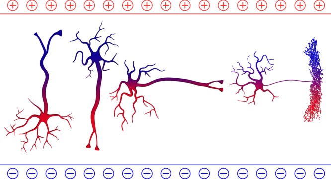
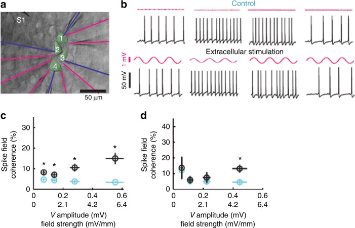
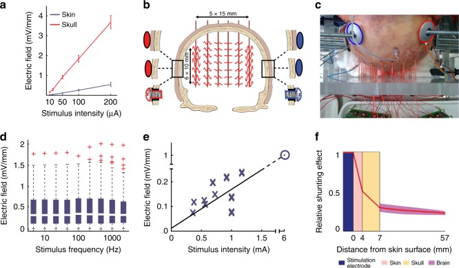
Noninvasive brain stimulation techniques are used in experimental and clinical fields for their potential effects on brain network dynamics and behavior. Transcranial electrical stimulation (TES), including transcranial direct current stimulation (tDCS) and transcranial alternating current stimulation (tACS), has gained popularity because of its convenience and potential as a chronic therapy. However, a mechanistic understanding of TES has lagged behind its widespread adoption. Here, we review data and modelling on the immediate neurophysiological effects of TES in vitro as well as in vivo in both humans and other animals. While it remains unclear how typical TES protocols affect neural activity, we propose that validated models of current flow should inform study design and artifacts should be carefully excluded during signal recording and analysis. Potential indirect effects of TES (e.g., peripheral stimulation) should be investigated in more detail and further explored in experimental designs. We also consider how novel technologies may stimulate the next generation of TES experiments and devices, thus enhancing validity, specificity, and reproducibility.


Conflict of interest statement

A.B. is the founder and owner of Amplipex and Neunos LLCs, which manufacture biosignal amplifiers and stimulator devices. A.B. and G.B. have filed a patent application about the ISP method. A.O. is an inventor on patents and patent applications describing methods and devices for noninvasive brain stimulation. B.K. is a paid consultant for Aqeel, LLC, which develops transcranial stimulation technology. L.P. has shares in Soterix Medical Inc., which develops transcranial stimulation technology. The remaining authors declare no competing interests.

Figures

Fig. 1
Fig. 1
The impact of orientation of neuronal compartments on TES-induced excitability. Four idealized neurons are shown with different orientation relative to the induced electric field. The electric field can affect the soma, dendrites, axon initial segment and the axon tree differently. The relationship between the electric field vector and the morphology/orientation of neurons and individual neuronal compartments determine whether the neuron will be net depolarized or hyperpolarized
Fig. 2
Fig. 2
Five postulated mechanisms to affect online spiking of neurons and networks patterns in response to different estimated magnitudes of TES. While the figure illustrates distinct effects, in reality the boundaries of mechanisms are blurred under most experimental conditions. Several mechanisms can act simultaneously in different networks of the same brain. The numbers on the vertical axis are merely estimates based on current data and theoretical considerations
Fig. 3
Fig. 3
Field-entrainment of spikes under idealized conditions in vitro. Synaptic transmission was blocked pharmacologically. a Four neurons with somata located within 100 μm of tissue were patched with intracellular electrodes (blue). Seven extracellular electrodes monitored extracellular voltage (Ve) fluctuations (magenta). An extracellular stimulation electrode (S1) was placed 50–80 μm from the recorded somata. b Each of the four intracellularly recorded neurons were depolarized to induced spiking. Spiking in the absence (top traces) and in the presence of extracellular stimulation (magenta; 100 nA at 1 Hz; bottom traces) is shown. c Spike field coherence (circles, mean; error bars, s.e.m.) between spikes and extracellular Ve is shown during 1 Hz extracellular stimulation (black) and control condition (cyan; circles indicate mean Ve amplitude at the soma and error bars indicate s.e.m.) as a function of stimulation strength. Asterisks indicate statistical significance of the spike-field difference between control and extracellular stimulation. d Spike-spike coordination (spike-field coherence for two simultaneously occurring spikes; essentially spike synchrony among neurons) during 1 Hz extracellular stimulation (black) and control (cyan) condition. Note that stronger fields are needed for coherent entrainment of neuronal spikes across the four neurons (d) than for inducing spike-field coherence for each neuron separately (c). Figure reproduced with permission
Fig. 4
Fig. 4
Current loss of TES in rodents and in human cadaver brains. a Compared to subcutaneous stimulation (red), transcutaneous stimulation (blue) generated several-fold weaker electric fields in a rodent model. b Schematic of the experimental arrangement for transcutaneous, subcutaneous, and epidural stimulation in cadavers, in a coronal plane. c Photograph of the cadaver skull with drilled holes and inserted matrix of recording electrodes. Stimulation electrodes, marked by blue and red circles for negative and positive polarity, respectively, were fixed to the skull. d Effect of stimulus frequency on intracerebral voltage gradients. Stimulus frequency between 5 and 1000 Hz has a minor effect on intracerebral gradients. e Extrapolation from measurements in human cadavers (blue crosses) suggests that approximately 6 mA current applied across the skin would induce 1 V/m intracerebral electric field (blue open circle). f Attenuation of charge flow (red line) through scalp (pink), skull (yellow), and brain (dark pink), as measured in human cadaver heads. Figure adapted from Figs. 1, 4, 5 in Voroslakos et al., (2018), under the Creative Commons License
Fig. 5
Fig. 5
Individualized models of transcranial electrical stimulation for two subjects with variable electrode placements. a Model of Subject 1, with 2 mA current injected at electrode Fp1 and return from electrode P3. b Model of Subject 2 with the same electrode configuration. c Model of Subject 2 with another electrode configuration attempting to achieve more focal stimulation. Note that stimulation is more focal but achieves weaker fields compared to b, due to increased current shunting through the skin between near-by electrodes. Electrode montages in b and c are obtained by a numerical optimization algorithm that attempts to achieve maximal intensity (b) or maximal focality (c) for the location indicated by a black circle. The current-flow models were generated from previously published data and methods. We used ROAST, a toolbox for realistic current-flow models of the human

References

    1. von Helmholtz, H. Treatise on Physiological Optics. Vol. III, (Dover Publications, New York, 1867).
    1. Rohracher, H. Ueber subjektive Lichterscheinungen bei Reizung mit Wechselströmen. Zeitschrift für Sinnesphysiologie66, 164–181 (1935).
    1. Barlow HB, Kohn HI, Walsh EG. Visual sensations aroused by magnetic fields. Am. J. Physiol. 1947;148:372–375. - PubMed
    1. Kar K, Krekelberg B. Transcranial electrical stimulation over visual cortex evokes phosphenes with a retinal origin. J. Neurophysiol. 2012;108:2173–2178. doi: 10.1152/jn.00505.2012. - DOI - PMC - PubMed
    1. Rudorfer, M., Henry, M. & Sackeim, H. in Psychiatry (eds. Tasman, A., Kay, J. & Lieberman, J.) 1539 (WB Saunders, Philadelphia, 1997).

Publication types