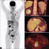
Fluroine-18 fluorodeoxyglucose-positron emission tomography/computer tomography in staging of renal cell carcinoma arising from a native kidney with liver and bone metastasis in a renal transplant patient
- PMID: 30505230
- PMCID: PMC6216737
- DOI: 10.4103/wjnm.WJNM_65_17
Fluroine-18 fluorodeoxyglucose-positron emission tomography/computer tomography in staging of renal cell carcinoma arising from a native kidney with liver and bone metastasis in a renal transplant patient
Abstract
Renal cell carcinoma (RCC) of the native kidney accounts for <5% of all malignancies found in transplant recipients. There have been only a few reported cases comprising of few renal transplant patients with RCC of native kidneys due to the relative rarity of the condition. Fluorine-18 Fluorodeoxyglucose (FDG)-positron emission tomography (PET)/computed tomography (CT) is used in the staging of RCC. Prognosis of metastatic RCC is poor. We report the first case of 55-year-old postrenal transplant recipient diagnosed with RCC of the native kidney with liver and bone metastases imaged using F-18 FDG PET/CT.
Keywords: Fluoride-18 Fluorodeoxyglucose-positron emission tomography/computed tomography; native kidney; renal cell carcinoma; renal transplant.
Conflict of interest statement
There are no conflicts of interest.
Figures

References
-
- Kasiske BL, Snyder JJ, Gilbertson DT, Wang C. Cancer after kidney transplantation in the United States. Am J Transplant. 2004;4:905–13. - PubMed
-
- Klatte T, Marberger M. Renal cell carcinoma of native kidneys in renal transplant patients. Curr Opin Urol. 2011;21:376–9. - PubMed
-
- Schwarz A, Vatandaslar S, Merkel S, Haller H. Renal cell carcinoma in transplant recipients with acquired cystic kidney disease. Clin J Am Soc Nephrol. 2007;2:750–6. - PubMed
-
- Goh A, Vathsala A. Native renal cysts and dialysis duration are risk factors for renal cell carcinoma in renal transplant recipients. Am J Transplant. 2011;11:86–92. - PubMed
Publication types
LinkOut - more resources
Full Text Sources

