

MicroRNA and Long Non-coding RNA Regulation in Skeletal Muscle From Growth to Old Age Shows Striking Dysregulation of the Callipyge Locus
- PMID: 30505320
- PMCID: PMC6250799
- DOI: 10.3389/fgene.2018.00548
MicroRNA and Long Non-coding RNA Regulation in Skeletal Muscle From Growth to Old Age Shows Striking Dysregulation of the Callipyge Locus
Abstract
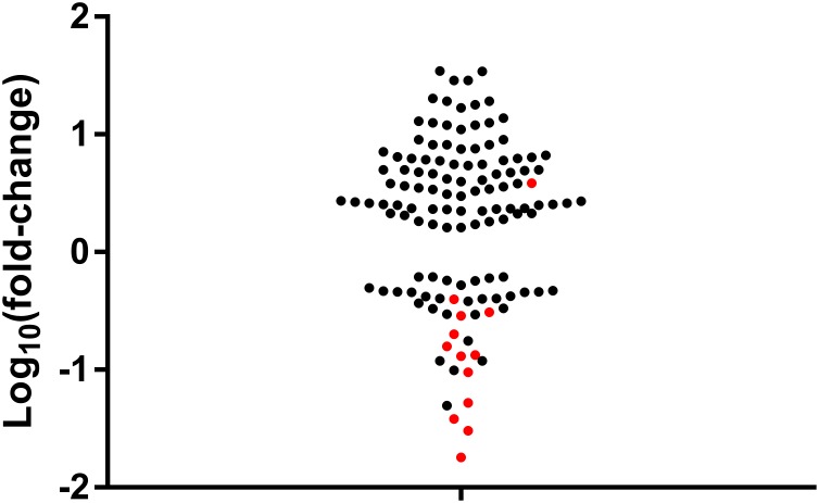
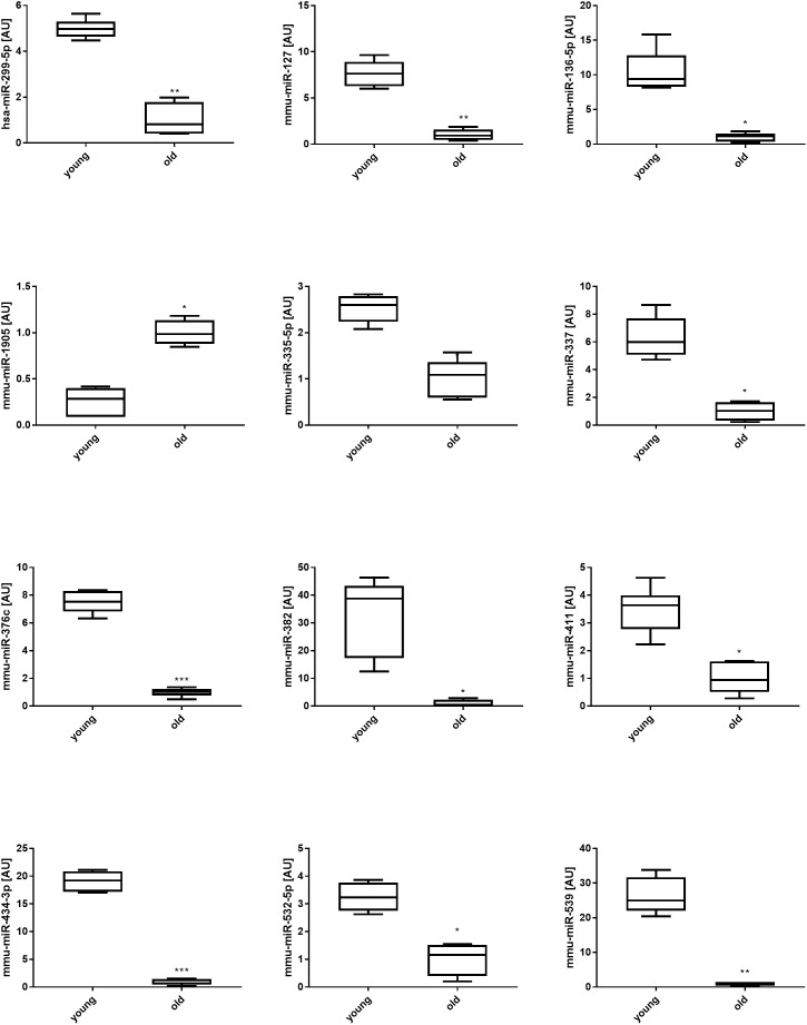
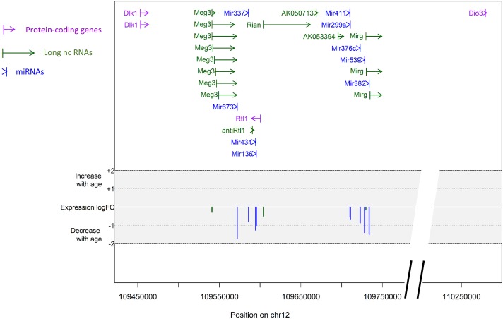
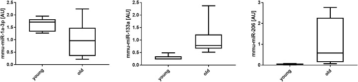
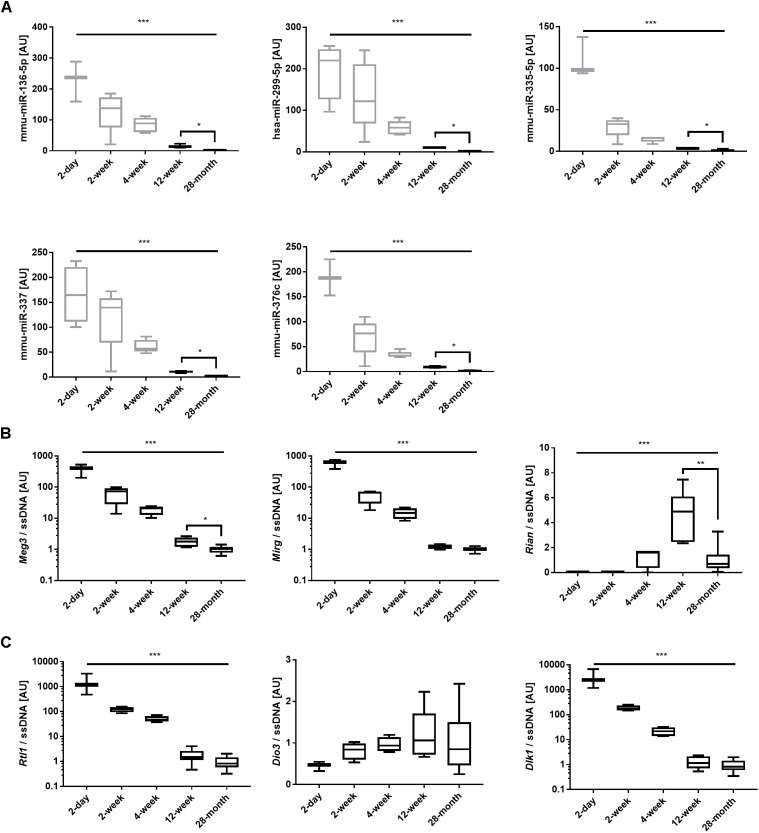
MicroRNAs (miRNAs) undergo high levels of regulation in skeletal muscle development and control skeletal muscle mass, function and metabolism over the lifespan. More recently, the role of long non-coding RNAs (lncRNAs) in skeletal muscle regulation has started to emerge. Following up on our recent study describing the expression pattern and putative roles of 768 miRNAs in the quadriceps muscle of mice at early life stages, we used a high-throughput miRNA qPCR-based array to assess the expression of the same miRNAs in 28-month old male mouse quadriceps muscle. In addition, we report the expression patterns of lncRNAs playing a putative role in muscle development and adaptation from growth to old age. Twelve miRNAs were significantly downregulated in 28-month old muscle when compared with 12-week old muscle. Ten of them clustered at the Dlk1-Dio3 locus, known as 'Callipyge,' which is associated with muscle development and hypertrophy. This collective downregulation was paralleled by decreases in the expression levels of the maternally expressed imprinted LncRNA coding genes Meg3 and Rian stemming from the same chromosomal region. In contrast, the paternally expressed imprinted Dlk1-Dio3 locus members Rtl1, Dio3, and Dlk1 and the muscle related lncRNAs lncMyoD1, Neat_v1, Neat_v2, and Malat1 underwent significant changes during growth, but their expression levels were not altered past the age of 12 weeks, suggesting roles limited to hyperplasia and early hypertrophy. In conclusion, collective muscle miRNA expression gradually decreases over the lifespan and a cluster of miRNAs and maternally expressed lncRNAs stemming from the Callipyge locus is significantly dysregulated in aging muscle. The Dlk1-Dio3 locus therefore represents a potential new mechanism for age-related muscle decline.
Keywords: aging; lncRNAs; miRNAs; sarcopenia; skeletal muscle.
Figures
Similar articles
-
Myostatin-deficiency in mice increases global gene expression at the Dlk1-Dio3 locus in the skeletal muscle.Oncotarget. 2017 Jan 24;8(4):5943-5953. doi: 10.18632/oncotarget.13966. Oncotarget. 2017. PMID: 27992376 Free PMC article.
-
Male-specific coordinated changes in expression of miRNA genes, but not other genes within the DLK1-DIO3 locus in multiple sclerosis.Gene. 2022 Aug 20;836:146676. doi: 10.1016/j.gene.2022.146676. Epub 2022 Jun 14. Gene. 2022. PMID: 35714798
-
Effect of DLK1 and RTL1 but not MEG3 or MEG8 on muscle gene expression in Callipyge lambs.PLoS One. 2009 Oct 9;4(10):e7399. doi: 10.1371/journal.pone.0007399. PLoS One. 2009. PMID: 19816583 Free PMC article.
-
Dysregulation of ncRNAs located at the DLK1‑DIO3 imprinted domain: involvement in urological cancers.Cancer Manag Res. 2019 Jan 15;11:777-787. doi: 10.2147/CMAR.S190764. eCollection 2019. Cancer Manag Res. 2019. PMID: 30697070 Free PMC article. Review.
-
A Hearty Dose of Noncoding RNAs: The Imprinted DLK1-DIO3 Locus in Cardiac Development and Disease.J Cardiovasc Dev Dis. 2018 Jul 10;5(3):37. doi: 10.3390/jcdd5030037. J Cardiovasc Dev Dis. 2018. PMID: 29996488 Free PMC article. Review.
Cited by
-
Long Non-coding RNA MALAT1 Is Depleted With Age in Skeletal Muscle in vivo and MALAT1 Silencing Increases Expression of TGF-β1 in vitro.Front Physiol. 2022 Jan 21;12:742004. doi: 10.3389/fphys.2021.742004. eCollection 2021. Front Physiol. 2022. PMID: 35126169 Free PMC article.
-
The Functional Role of Long Non-Coding RNA in Myogenesis and Skeletal Muscle Atrophy.Cells. 2022 Jul 25;11(15):2291. doi: 10.3390/cells11152291. Cells. 2022. PMID: 35892588 Free PMC article. Review.
-
The super-healing MRL strain promotes muscle growth in muscular dystrophy through a regenerative extracellular matrix.JCI Insight. 2024 Jan 4;9(3):e173246. doi: 10.1172/jci.insight.173246. JCI Insight. 2024. PMID: 38175727 Free PMC article.
-
Competing endogenous network analysis identifies lncRNA Meg3 activates inflammatory damage in UVB induced murine skin lesion by sponging miR-93-5p/epiregulin axis.Aging (Albany NY). 2019 Nov 24;11(22):10664-10683. doi: 10.18632/aging.102483. Epub 2019 Nov 24. Aging (Albany NY). 2019. PMID: 31761787 Free PMC article.
-
A Meta-Analysis of Brain DNA Methylation Across Sex, Age, and Alzheimer's Disease Points for Accelerated Epigenetic Aging in Neurodegeneration.Front Aging Neurosci. 2021 Mar 11;13:639428. doi: 10.3389/fnagi.2021.639428. eCollection 2021. Front Aging Neurosci. 2021. PMID: 33790779 Free PMC article.
References
-
- Barns M., Gondro C., Tellam R. L., Radley-Crabb H. G., Grounds M. D., Shavlakadze T. (2014). Molecular analyses provide insight into mechanisms underlying sarcopenia and myofibre denervation in old skeletal muscles of mice. Int. J. Biochem. Cell Biol. 53 174–185. 10.1016/j.biocel.2014.04.025 - DOI - PubMed
LinkOut - more resources
Full Text Sources
Miscellaneous