. 2018 Nov 26:6:79.
doi: 10.1186/s40560-018-0348-y. eCollection 2018.

Circulating histone H3 levels are increased in septic mice in a neutrophil-dependent manner: preclinical evaluation of a novel sandwich ELISA for histone H3

Affiliations

Circulating histone H3 levels are increased in septic mice in a neutrophil-dependent manner: preclinical evaluation of a novel sandwich ELISA for histone H3

Takashi Ito et al. J Intensive Care. .

Abstract

Background: Nuclear histone proteins are released into the extracellular space during sepsis and act as major mediators of death. However, circulating histone levels have not been precisely quantified.

Methods: We developed a novel enzyme-linked immunosorbent assay (ELISA) for detection of circulating histone H3 levels and evaluated its performance. Using the ELISA, we measured plasma histone H3 levels in C57BL/6 J mice subjected to cecal ligation and puncture (CLP)-induced sepsis.

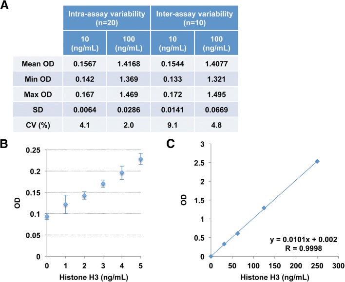
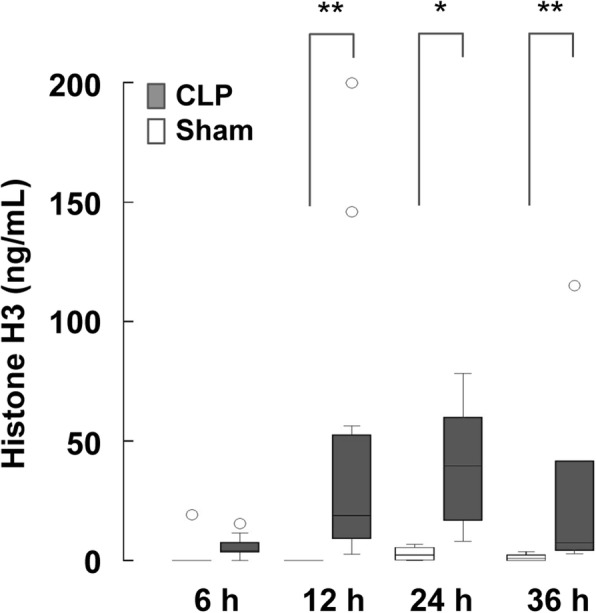
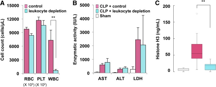
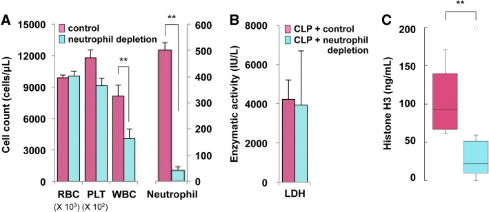
Results: The newly developed ELISA enabled reproducible measurement of histone H3 levels with a working range up to 250 ng/mL. Using the ELISA, we found that plasma histone H3 levels were elevated in septic mice compared with sham-operated mice (p < 0.01). The elevation of histone H3 levels was abrogated when neutrophils were depleted (p < 0.01).

Conclusions: Our novel ELISA provides reproducible measurements of histone H3 levels. Circulating histone H3 levels are increased in septic mice in a neutrophil-dependent manner. Further studies are needed to evaluate the clinical utility of histone H3 levels in patients with sepsis.

Keywords: ELISA; Histone; Neutrophil; Sepsis.


Conflict of interest statement

All experiments involving animals were approved by the Institutional Animal Care and Use Committee of Kagoshima University.Not applicable.The ELISA for histone H3 is a product in development of Shino-Test Corporation where SO and SY are employed. IM holds an endowed chair at Kagoshima University and received a research fund from Shino-Test Corporation. The fund is for academic promotion and is not directly related to this study. The remaining authors declare that they have no competing interests.Springer Nature remains neutral with regard to jurisdictional claims in published maps and institutional affiliations.

Figures

Fig. 1
Fig. 1
Validation of the newly developed ELISA for detection of circulating histone H3 levels. a Intra-assay and inter-assay precisions were analyzed using serum samples containing histone H3 at concentrations of 10 and 100 ng/mL. The intra-assay (n = 20) and inter-assay (n = 10) CVs were 2.0–4.1% and 4.8–9.1%, respectively. OD, optical density; CV, coefficient of variation. b The detection limit determined by the mean ± 2.6 SD method was 2 ng/mL. c Linearity was observed in the range up to 250 ng/mL
Fig. 2
Fig. 2
Circulating histone H3 levels are elevated in mice with CLP. Serum histone H3 levels in C57BL/6 J mice with CLP-induced sepsis or sham operation are shown. Serum samples were collected at 6, 12, 24, and 36 h after CLP (n = 6–12 per group) or sham operation (n = 4–10 per group). Representative data of two or more independent experiments are shown. Differences in circulating histone H3 levels between CLP mice and sham mice were analyzed by the Mann–Whitney U test. *p < 0.05, **p < 0.01
Fig. 3
Fig. 3
Circulating histone H3 levels are mainly derived from leukocytes in CLP mice. a Leukocyte depletion was achieved by intraperitoneal injection of 150 mg/kg and 100 mg/kg cyclophosphamide at 72 h and 24 h prior to blood cell counts, respectively. RBC, red blood cell (× 103 cells/μL); PLT, platelet (× 102 cells/μL); WBC, white blood cell (cells/μL). Differences in WBC counts between cyclophosphamide-injected mice (leukocyte depletion, n = 5, mean ± SD) and saline-injected mice (control, n = 6, mean ± SD) were analyzed by Welch’s t-test. **p < 0.01. b Leukocyte depletion was conducted by intraperitoneal injection of 150 mg/kg and 100 mg/kg cyclophosphamide at 72 and 24 h prior to CLP, respectively. For assessment of cellular damage, the activities of AST, ALT, and LDH at 24 h after CLP or sham operation were examined. The AST, ALT, and LDH levels at 24 h after CLP did not differ significantly between leukocyte-depleted mice (n = 9, mean ± SD) and control mice (n = 10, mean ± SD). c Serum histone H3 levels at 24 h after CLP were examined in leukocyte-depleted mice (n = 9) and control mice (n = 10). Representative data of two independent experiments are shown. Differences in circulating histone H3 levels were analyzed by the Mann–Whitney U test. **p < 0.01
Fig. 4
Fig. 4
Circulating histone H3 levels are mainly derived from neutrophils in CLP mice. a Neutrophil depletion was conducted by intravenous injection of 100 μg/mouse of anti-Ly-6G antibody at 72 and 24 h prior to blood cell counts. RBC, red blood cell (× 103 cells/μL); PLT, platelet (× 102 cells/μL); WBC, white blood cell (cells/μL). Differences in white blood cell (WBC) and neutrophil counts between anti-Ly-6G antibody-injected mice (neutrophil depletion, n = 8, mean ± SD) and isotype control antibody-injected mice (control, n = 5, mean ± SD) were analyzed by Welch’s t test. **p < 0.01. b Neutrophil depletion was achieved by intravenous injection of 100 μg/mouse of anti-Ly-6G antibody at 72 and 24 h prior to CLP. For assessment of cellular damage, the serum LDH levels at 24 h after CLP were examined. The serum LDH levels at 24 h after CLP did not differ significantly between neutrophil-depleted mice (n = 6, mean ± SD) and control mice (n = 10, mean ± SD). c Serum histone H3 levels at 24 h after CLP were examined in neutrophil-depleted mice (n = 8) and control mice (n = 10). Representative data of two independent experiments are shown. Differences in circulating histone H3 levels were analyzed by the Mann–Whitney U test. **p < 0.01

References

    1. Cohen J. The immunopathogenesis of sepsis. Nature. 2002;420(6917):885–891. doi: 10.1038/nature01326. - DOI - PubMed
    1. Singer M, Deutschman CS, Seymour CW, Shankar-Hari M, Annane D, Bauer M, Bellomo R, Bernard GR, Chiche JD, Coopersmith CM, et al. The third international consensus definitions for sepsis and septic shock (Sepsis-3) JAMA. 2016;315(8):801–810. doi: 10.1001/jama.2016.0287. - DOI - PMC - PubMed
    1. Hotchkiss RS, Karl IE. The pathophysiology and treatment of sepsis. N Engl J Med. 2003;348(2):138–150. doi: 10.1056/NEJMra021333. - DOI - PubMed
    1. Wang H, Bloom O, Zhang M, Vishnubhakat JM, Ombrellino M, Che J, Frazier A, Yang H, Ivanova S, Borovikova L, et al. HMG-1 as a late mediator of endotoxin lethality in mice. Science. 1999;285(5425):248–251. doi: 10.1126/science.285.5425.248. - DOI - PubMed
    1. Xu J, Zhang X, Pelayo R, Monestier M, Ammollo CT, Semeraro F, Taylor FB, Esmon NL, Lupu F, Esmon CT. Extracellular histones are major mediators of death in sepsis. Nat Med. 2009;15(11):1318–1321. doi: 10.1038/nm.2053. - DOI - PMC - PubMed

LinkOut - more resources