

Ontogeny and phylogeny of the mammalian chondrocranium: the cupula nasi anterior and associated structures of the anterior head region
- PMID: 30505462
- PMCID: PMC6260904
- DOI: 10.1186/s40851-018-0112-0
Ontogeny and phylogeny of the mammalian chondrocranium: the cupula nasi anterior and associated structures of the anterior head region
Abstract
Background: The study of chondrocrania has a long tradition with a focus on single specimens and stages. It revealed great interspecific diversity and a notion of intraspecific variation. As an embryonic structure, the chondrocranium is subject to major changes in ontogeny with resorption and ossification of different cartilaginous structures. The cupula nasi anterior is the anteriormost portion of the cartilaginous nasal capsule and is expected to mirror much of the animal's life history and lifestyle. Its diversity in mammals is reflected in the external nasal anatomy of newborns. Marsupials and placentals show marked differences, likely related to breathing and suckling behavior.
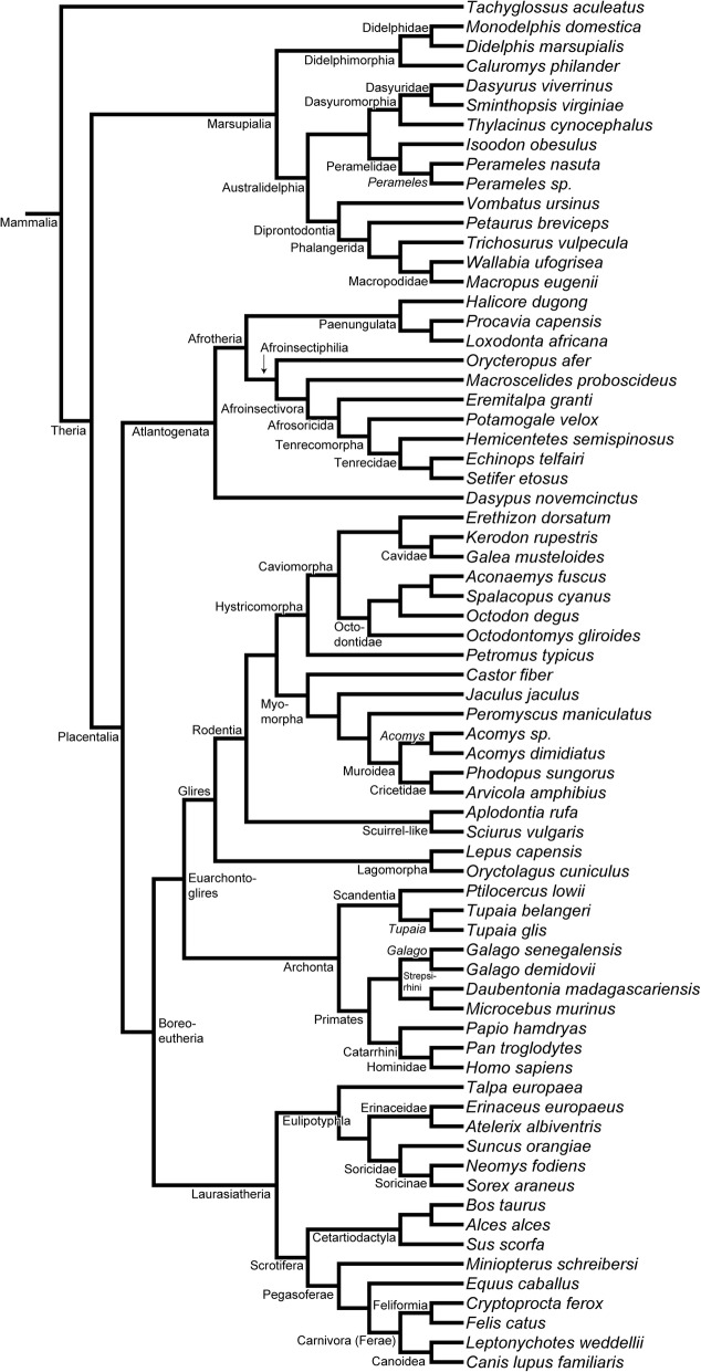
Results: We examined histological sections of five marsupial and three placentals species and traced the development of the cupula nasi anterior and the anterior nasal capsule. We found ontogenetic variation for nearly 50% of the 43 characters defined herein. By comparing to the literature and considering ontogenetic variation, we performed an analysis of character evolution in 70 mammalian species and reconstructed the nasal anatomy of the therian ancestor.
Conclusions: At birth, marsupials have a complete but simple cupula nasi anterior, whereas placentals display a more diverse morphology due to reductions and variations of chondrocranial elements. The more compact nasal capsule in marsupials is related to a long and strong fixation to the mother's teat after birth. Within marsupials and placentals, several derived characters distinguish major taxa, probably related to developmental and functional constraints. The reconstructed ancestral anatomy of the cupula nasi anterior supports the hypothesis that the therian ancestor was placental-like and that the marsupial lifestyle is more derived.
Keywords: Chondrocranium; Cupula nasi anterior; Mammalia; Ontogeny; Therian ancestor.
Conflict of interest statement
Not applicable.Not applicable.The authors declare that they have no competing interests.Springer Nature remains neutral with regard to jurisdictional claims in published maps and institutional affiliations.
Figures
Similar articles
-
Development of the cetacean nasal skull.Adv Anat Embryol Cell Biol. 1999;149:1-143. doi: 10.1007/978-3-642-58612-5. Adv Anat Embryol Cell Biol. 1999. PMID: 10091359
-
On the development of the chondrocranium and the histological anatomy of the head in perinatal stages of marsupial mammals.Zoological Lett. 2017 Feb 12;3:1. doi: 10.1186/s40851-017-0062-y. eCollection 2017. Zoological Lett. 2017. PMID: 28203388 Free PMC article. Review.
-
Ossification heterochrony in the therian postcranial skeleton and the marsupial-placental dichotomy.Evolution. 2008 Aug;62(8):2027-41. doi: 10.1111/j.1558-5646.2008.00424.x. Epub 2008 May 16. Evolution. 2008. PMID: 18489720
-
Pedomorphosis in the ancestry of marsupial mammals.Curr Biol. 2023 Jun 5;33(11):2136-2150.e4. doi: 10.1016/j.cub.2023.04.009. Epub 2023 Apr 28. Curr Biol. 2023. PMID: 37119816
-
Ontogenetic transformation of the cartilaginous nasal capsule in mammals, a review with new observations on bats.Anat Rec (Hoboken). 2025 Jul;308(7):1867-1883. doi: 10.1002/ar.25152. Epub 2023 Jan 16. Anat Rec (Hoboken). 2025. PMID: 36647334 Review.
Cited by
-
It takes two: Building the vertebrate skull from chondrocranium and dermatocranium.Vertebr Zool. 2020 Apr;70(4):587-600. Epub 2020 Oct 28. Vertebr Zool. 2020. PMID: 33163116 Free PMC article.
-
Evolution, conservatism and overlooked homologies of the mammalian skull.Philos Trans R Soc Lond B Biol Sci. 2023 Jul 3;378(1880):20220081. doi: 10.1098/rstb.2022.0081. Epub 2023 May 15. Philos Trans R Soc Lond B Biol Sci. 2023. PMID: 37183902 Free PMC article. Review.
-
Postnatal development in a marsupial model, the fat-tailed dunnart (Sminthopsis crassicaudata; Dasyuromorphia: Dasyuridae).Commun Biol. 2021 Sep 2;4(1):1028. doi: 10.1038/s42003-021-02506-2. Commun Biol. 2021. PMID: 34475507 Free PMC article.
References
-
- Kaucka M, Adameyko I. Evolution and development of the cartilaginous skull: from a lancelet towards a human face. Seminars in Cell & Developmental Biology; in press. - PubMed
-
- Maier W. Ontogeny of the Nasal Capsule in Cercopithecoids: a Contribution to the Comparative and Evolutionary Morphology of Catarrhines. In: Old World Monkeys. Cambridge: Cambridge University Press; 2000. p. 99–132.
-
- Maier W. Zur evolutiven und funktionellen Morphologie des Gesichtsschädels der Primaten. Z Morphol Anthropol. 1993;79:279–299. - PubMed
-
- Novacek MJ. Patterns of Diversity in the Mammalian Skull. The Skull. 1993;2:438–545.
LinkOut - more resources
Full Text Sources
Research Materials
