

Surgery for parasitic lung infestations: roles in diagnosis and treatment
- PMID: 30505532
- PMCID: PMC6218367
- DOI: 10.21037/jtd.2018.08.32
Surgery for parasitic lung infestations: roles in diagnosis and treatment
Abstract
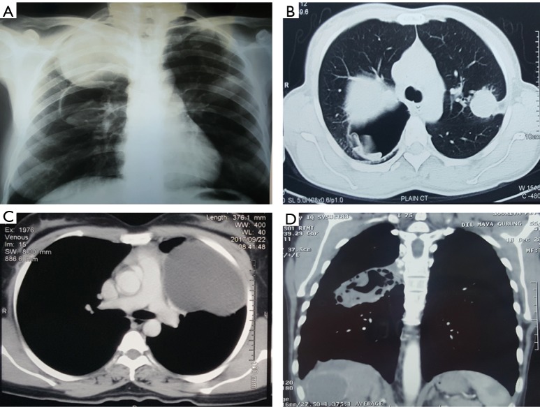
Pulmonary parasitic infestations are a worldwide problem associated with significant morbidity and socioeconomic impact. They are known to have varied clinical presentations and radiological appearances. Prevention of parasite transmission and medical treatment of cases form the two pillars of control of these diseases. The role of surgery is limited to the diagnosis and definitive treatment of the minority of pulmonary parasitic afflictions, most notably hydatidosis. Despite surgery being established as the treatment of choice in pulmonary hydatid cysts (PHCs) for over half a century, variations and unresolved controversies persist regarding the best surgical technique. Complications brought on by cyst rupture, multiplicity and multi-organ involvement add complexity to treatment decisions. The development of video-assisted thoracoscopic surgery (VATS) brings the promise of reduced peri-operative morbidity but is yet to be universally accepted as a safe technique. In this review, we endeavor to discuss the common pulmonary infestations focusing on the current trends and controversies surrounding surgery for PHC.
Keywords: Echinococcosis; pulmonary parasitic infestations; surgical management.
Conflict of interest statement
Conflicts of Interest: The authors have no conflicts of interest to declare.
Figures

Similar articles
-
[Complicated hydatid cysts of the lung].Rev Mal Respir. 2009 Sep;26(7):727-34. doi: 10.1016/s0761-8425(09)72423-4. Rev Mal Respir. 2009. PMID: 19953014 French.
-
Therapeutic evaluation of video-assisted thoracoscopic surgery versus open thoracotomy for pediatric pulmonary hydatid disease.J Cardiothorac Surg. 2016 Aug 5;11(1):129. doi: 10.1186/s13019-016-0525-9. J Cardiothorac Surg. 2016. PMID: 27495934 Free PMC article.
-
Video-assisted Thoracoscopic Surgery (VATS) with mini-thoracotomy for the management of pulmonary hydatid cysts.J Cardiothorac Surg. 2018 May 2;13(1):35. doi: 10.1186/s13019-018-0716-7. J Cardiothorac Surg. 2018. PMID: 29716636 Free PMC article.
-
Pulmonary hydatid and other lung parasitic infections.Curr Opin Pulm Med. 2002 May;8(3):218-23. doi: 10.1097/00063198-200205000-00012. Curr Opin Pulm Med. 2002. PMID: 11981312 Review.
-
Cystic pulmonary hydatidosis.Lung India. 2016 Mar-Apr;33(2):179-91. doi: 10.4103/0970-2113.177449. Lung India. 2016. PMID: 27051107 Free PMC article. Review.
Cited by
-
Current Management of Pulmonary Hydatid Cyst.Eurasian J Med. 2025 Feb 3;57(1):1-7. doi: 10.5152/eurasianjmed.2025.24761. Eurasian J Med. 2025. PMID: 40377486 Free PMC article.
-
Hydatid Cyst or Echinococcosis: A Comprehensive Review of Transmission, Clinical Manifestations, Diagnosis, and Multidisciplinary Treatment.Cureus. 2024 Jul 2;16(7):e63713. doi: 10.7759/cureus.63713. eCollection 2024 Jul. Cureus. 2024. PMID: 39099980 Free PMC article. Review.
-
Modified capitonnage technique for giant pulmonary hydatid cyst surgery.Interact Cardiovasc Thorac Surg. 2021 Oct 29;33(5):721-726. doi: 10.1093/icvts/ivab152. Interact Cardiovasc Thorac Surg. 2021. PMID: 34041544 Free PMC article.
-
Comparing Capitonnage and Uncapitonnage Techniques for Pulmonary Hydatid Cysts: A Systematic Review and Meta-analysis.Eurasian J Med. 2023 Nov 2;55(Suppl 1):S35-42. doi: 10.5152/eurasianjmed.2023.22281. Online ahead of print. Eurasian J Med. 2023. PMID: 37916996 Free PMC article.
-
Surgical management of pulmonary hydatid cysts in children in KwaZulu-Natal Province, South Africa.Afr J Thorac Crit Care Med. 2020 Oct 13;26(3):10.7196/AJTCCM.2020.v26i3.108. doi: 10.7196/AJTCCM.2020.v26i3.108. eCollection 2020. Afr J Thorac Crit Care Med. 2020. PMID: 34240027 Free PMC article.
References
Publication types
LinkOut - more resources
Full Text Sources
Molecular Biology Databases