. 2017 Nov;1(11):862-877.
doi: 10.1038/s41551-017-0154-1. Epub 2017 Nov 10.

Glial responses to implanted electrodes in the brain

Affiliations

Glial responses to implanted electrodes in the brain

Joseph W Salatino et al. Nat Biomed Eng. 2017 Nov.

Erratum in

Abstract

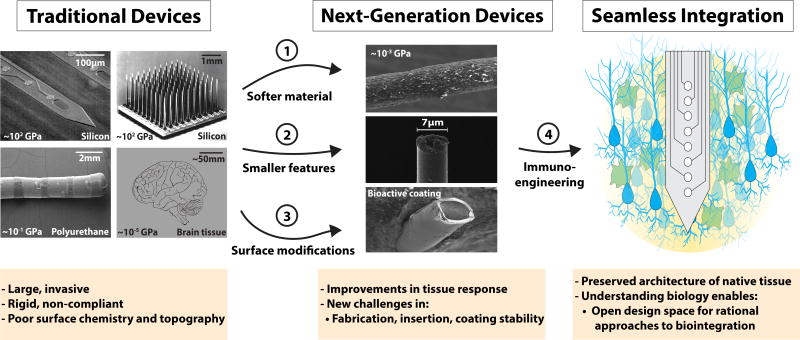
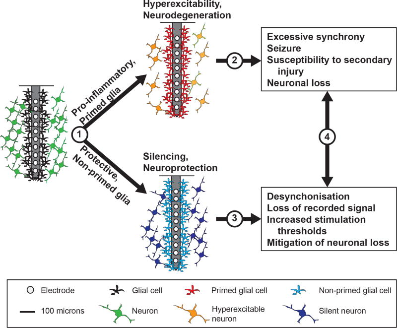
The use of implants that can electrically stimulate or record electrophysiological or neurochemical activity in nervous tissue is rapidly expanding. Despite remarkable results in clinical studies and increasing market approvals, the mechanisms underlying the therapeutic effects of neuroprosthetic and neuromodulation devices, as well as their side effects and reasons for their failure, remain poorly understood. A major assumption has been that the signal-generating neurons are the only important target cells of neural-interface technologies. However, recent evidence indicates that the supporting glial cells remodel the structure and function of neuronal networks and are an effector of stimulation-based therapy. Here, we reframe the traditional view of glia as a passive barrier, and discuss their role as an active determinant of the outcomes of device implantation. We also discuss the implications that this has on the development of bioelectronic medical devices.


Conflict of interest statement

Competing interests The authors declare no competing financial interests.

Figures

Fig. 1
Fig. 1
Traditional electrode arrays incite gliosis. af, Devices (ac) are shown above the associated histology images (df). a, Michigan-style array. b, Utah-style array. c, DBS lead. d, Rat histology from a Michigan-style multielectrode array (four weeks), with labelled astrocytes (GFAP, green) and microglia (ED1, red). e, Histology from a primate with Utah array implanted, with microglia labelled (IBA1, red), at 17 weeks. f, Human DBS lead implant at ~38 months, with labelled astrocytes (GFAP, magenta; white arrowheads), microglia (IBA1, cyan; white arrows) and all cell nuclei (CyQUANT, yellow). Scales bars: a,d,f, 100 µm; b, 1 mm; c, 2 mm; e, 28 µm. The asterisks in df indicate injury. ED1, antibody to cluster of differentiation 68; IBA1, ionized calcium binding adaptor molecule 1. Figure reproduced from: a, ref., IEEE; b, ref., Elsevier; c, ref., Oxford Univ. Press; d, ref., Elsevier; e, ref., IOP Publishing; f, ref., Springer.
Fig. 2
Fig. 2
In vivo multiphoton imaging of the glial response to the implantation of a multielectrode array. Astrocytes and oligodendrocytes (sulfarhodamine, false-coloured purple in a), neurovasculature (intravascular sulfarhodamine, red in all panels) and microglia (the transgenic line CX3CR1-GFP, green in all panels) are shown. a, Microglia display an amoeboid morphology and encapsulate two shanks of a 4 × 4 NeuroNexus array six hours following implantation. b, Microglia form a compact scar around two shanks of a 1 × 3 Blackrock array at two months post-implantation. c, Microglia activation and lamellipodia ensheathment of an implanted silicon/silicon-oxide microelectrode. d, Microglia avoid the silicon/silicon-oxide microelectrode surface when covalently coated with neurocamouflage protein L1CAM. Scale bars, 100 µm. Figure adapted from: a, ref., IOP Publishing; b, ref., Elsevier; c,d, ref., Elsevier.
Fig.3
Fig.3
Evidence for a negative impact of increased gliosis on recording quality. ad, Representative images from four animals demonstrate the range of endpoint histological outcomes (from ‘good’ to ‘poor’, left to right). The figure has been generated after additional analysis on data collected in a previous study. Neuronal nuclei (NeuN, green) and astrocytes (GFAP, red) surrounding probe tracts are shown, and the associated average neuronal and non- neuronal density data are listed (area binned cell counts, neuronal density (ND) and non-neuronal density (NND), in cells mm−2). Recording segments with signal-to-noise-ratio (SNR) values representative of the average value for each animal are depicted (the SNRs calculated from peak-to-peak noise result in lower values than those calculated from root-mean-square noise),. Recording quality improved with decreased NND and increased ND/NND (P < 0.05, Spearman’s ρ, n = 6). Impedance increased with increased NND (P < 0.05, Spearman’s ρ, n = 6). Animals in a and c were drug-treated while b and d correspond to the controls. Scale bar, 100 µm. Figure adapted from ref., Elsevier.
Fig.4
Fig.4
Potential mechanisms of the active modulation of neurotransmission by glia. a, Insertional trauma incites reactive gliosis and impacts neuronal function through modifications to the local neurochemical environment. Punctured cellular membranes release ATP into the local extracellular space, whereby activated microglia and astrocytes are recruited to release glutamate, cytokines and ATP. The resulting signalling cascades ultimately reinforce reactive gliosis and impact local neuronal health and function. The dashed box indicates the region of synaptic silencing depicted in b. Neuronal excitotoxicity is another potential consequence of reactive signalling. b, As injured cells and reactive microglia release excess ATP, activated astrocytes are able to silence neuronal activity through two synaptic mechanisms. (1) Glutamate and ATP release, which generate a positive-feedback loop; ATP is rapidly hydrolysed to adenosine in the synapse, where adenosine is able to act on presynaptic A1Rs to inhibit Ca2+ channels and prevent vesicle release (presynaptic silencing), and to act on postsynaptic A1Rs to open K+ and Cl channels and prevent the generation of action potentials (postsynaptic silencing). (2) TSP production and release, which forms ultrastructurally normal, but functionally silent synapses. These postsynaptic terminals lack AMPARs, which are required to alleviate the Mg2+ block on NMDARs, therefore preventing effective signal transfer from the presynapse (postsynaptic silencing). A1R, adenosine A1 receptor; P2R, purinergic P2 receptor; GluT, glutamate transporter.
Fig.5
Fig.5
Next-generation arrays mitigate gliosis. af, Devices (ac) are shown above the associated histology images (df). a, A mechanically adaptive nanocomposite microelectrode becomes compliant on implantation. b, A hollow-architecture parylene-based microelectrode places sites away from the stiff penetrating shaft, along 4-µm-wide lateral support arms. c, A syringe-injectable mesh electronics mimics brain parenchyma with sites featured along an interwoven structure. d, Astrocytes labelled (GFAP, green) around mechanically compliant probe at eight weeks. e, Astrocytes (GFAP, red), microglia (OX42, green), and all cells (Hoechst, blue) labelled around the stiff electrode-penetrating shaft (S) and lateral edge (L) at four weeks. f, Astrocytes labelled (GFAP, cyan) around a syringe-injected mesh (blue) at one year. Scale bars: a, 500 µm; b,d,f, 100 µm; c, 250 µm; e, 50 µm. Figure reproduced from: a, ref., IOP Publishing; b,e, ref., Elsevier; c, ref., American Chemical Society; d, ref., IOP Publishing; f, ref., Nature America Inc.
Fig.6
Fig.6
Opportunities for further enquiry in device design. Future work will need to uncover the effects of electrode properties on the molecular pathways that shape gliosis, including: (1) the degree of softness and corresponding inflammation from mechanoactivation of glia, and the evolution of the effect on gliosis over time (such as mechanical mismatch, micromotion, and the state of glial reactivity and ‘priming’); (2) the relationship between feature size and architecture on inciting and priming inflammatory gliosis around the injury, and the evaluation of the long-term consequences (such as hyperexcitability, excitotoxicity and degeneration) on device function; (3) the effects of surface modifications (chemistry and topography) on shaping reactive signalling at the interface (receptor activation and cytokine/gliotransmitter release) and the corresponding consequences on recording and stimulation performance; (4) targeted approaches to modify immune responses will need to be incorporated to achieve seamless integration, which should be guided by their impact on glial signalling, reactivity and device performance. Traditional devices images reproduced from refs (see Fig. 1 for credits). Next-generation devices reproduced from: top and bottom, ref., RSC; middle, ref., Macmillan Publishers Ltd.
Fig.7
Fig.7
Opportunities for further biological enquiry. (1) The factors responsible for the ‘tipping point’ between reactive and non-reactive glial states, and the implications of glial priming on the safety of high-density arrays and of multiple implant strategies. (2) The contribution of hyperexcitability to neuronal loss and recorded signal quality, and the underlying relationship with a primed glial state. (3) Glial-mediated neuronal silencing surrounding implants and the relationship to recorded signals and stimulation thresholds. (4) The relationship between device performance and the time course of glial effects, for insights into the sources of performance variability, plasticity and placebo effects of device insertion, as well as therapeutic effects and side effects in a broad range of device applications.

References

    1. Kasthuri N, Lichtman JW. Neurocartography. Neuropsychopharmacology. 2010;35:342–343. - PMC - PubMed
    1. Oberlaender M, et al. Three-dimensional axon morphologies of individual layer 5 neurons indicate cell type-specific intracortical pathways for whisker motion and touch. Proc. Natl Acad. Sci USA. 2011;108:4188–4193. - PMC - PubMed
    1. Kubota Y. Untangling GABAergic wiring in the cortical microcircuit. Curr. Opin. Neurobiol. 2014;26:7–14. - PubMed
    1. Zhang Y, Barres BA. Astrocyte heterogeneity: an underappreciated topic in neurobiology. Curr. Opin. Neurobiol. 2010;20:588–594. - PubMed
    1. Miocinovic S, Somayajula S, Chitnis S, Vitek JL. History, applications, and mechanisms of deep brain stimulation. JAMA Neurol. 2013;70:163–171. - PubMed