

Optical Rehabilitation of a Patient with Keratoconus and Nystagmus
- PMID: 30505870
- PMCID: PMC6229678
Optical Rehabilitation of a Patient with Keratoconus and Nystagmus
Abstract
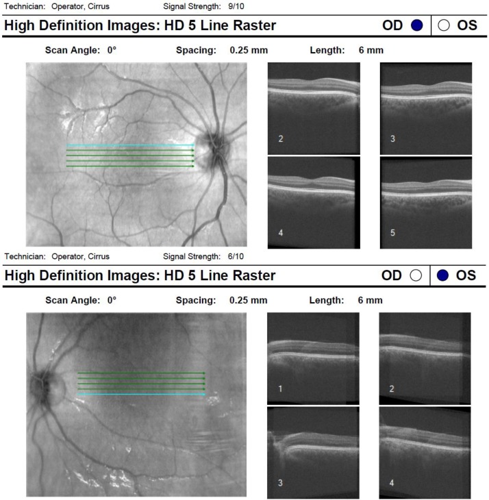
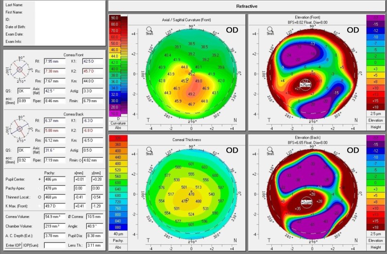
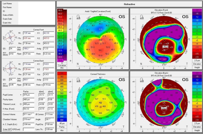
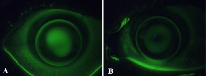
Keratoconus is a progressive corneal disease characterized by bilateral yet usually asymmetric thinning of the cornea with an onset typically in teenage years. While it often presents as an isolated condition, keratoconus may also be associated with many systemic and/or ocular diseases, such as connective tissue and chromosomal disorders. Its association with nystagmus has been described in Leber's congenital amaurosis, where patients also exhibit abnormal pupillary responses, early-onset retinal dystrophy, mental developmental delays, and eventual blindness. The case described here, however, was a high-functioning teenager with keratoconus and infantile nystagmus, and oscillopsia on left gaze and a compensatory head turn to the patient's left. The initial distance visual acuities of 20/60 and 20/150 in the right and left eye, respectively improved to 20/25 and 20/40 by the use of corneal rigid gas permeable contact lenses. In addition, the patient's neck strain and overall gait were eased by yoked prism spectacles.
Keywords: Ectasia; Infantile Nystagmus; Keratoconus; Rigid Contact Lens; Yoked Prisms.
Conflict of interest statement
Ethical issues have been completely observed by the authors. All named authors meet the International Committee of Medical Journal Editors (ICMJE) criteria for authorship of this manuscript, take responsibility for the integrity of the work as a whole, and have given final approval for the version to be published. No conflict of interest has been presented.
Figures

Similar articles
-
Optical quality and visual performance with customised soft contact lenses for keratoconus.Ophthalmic Physiol Opt. 2014 Sep;34(5):528-39. doi: 10.1111/opo.12133. Epub 2014 Apr 24. Ophthalmic Physiol Opt. 2014. PMID: 24758229
-
Contact lenses as the best conservative treatment of newly diagnosed keratoconus--epidemiological retrospective study.Coll Antropol. 2014 Dec;38(4):1115-8. Coll Antropol. 2014. PMID: 25842743
-
Managing keratoconus with reverse-geometry and dual-geometry contact lenses: a case report.Eye Contact Lens. 2008 Jan;34(1):71-5. doi: 10.1097/ICL.0b013e31805e35fa. Eye Contact Lens. 2008. PMID: 18180689
-
[Keratoconus].J Fr Ophtalmol. 2013 Sep;36(7):618-26. doi: 10.1016/j.jfo.2013.05.004. Epub 2013 Jul 30. J Fr Ophtalmol. 2013. PMID: 23911067 Review. French.
-
Contact lens management of keratoconus.Clin Exp Optom. 2015 Jul;98(4):299-311. doi: 10.1111/cxo.12300. Clin Exp Optom. 2015. PMID: 26104589 Review.
References
-
- Zadnik K, Barr JT, Gordon MO, Edrington TB. Biomicroscopic signs and disease severity in keratoconus Collaborative Longitudinal Evaluation of Keratoconus (CLEK) Study Group. Cornea. 1996;15(2):139–46. pmid: 8925661. - PubMed
-
- Zadnik K, Steger-May K, Fink BA, Joslin CE, Nichols JJ, Rosenstiel CE, et al. Between-eye asymmetry in keratoconus. Cornea. 2002;21(7):671–9. pmid: 12352084. - PubMed
-
- Krachmer JH, Feder RS, Belin MW. Keratoconus and related noninflammatory corneal thinning disorders. Surv Ophthalmol. 1984;28(4):293–322. - PubMed
-
- Robertson I. Keratoconus and the Ehlers-Danlos syndrome: a new aspect of keratoconus. Med J Aust. 1975;1(18):571–3. pmid: 1143149. - PubMed
Publication types
LinkOut - more resources
Full Text Sources