Review
. 2018 Oct 31:3:86.
doi: 10.21037/tgh.2018.10.07. eCollection 2018.

Incisions and reconstruction approaches for large sarcomas

Affiliations
Review

Incisions and reconstruction approaches for large sarcomas

Leigh J Spera et al. Transl Gastroenterol Hepatol. .

Abstract

Large intraabdominal, retroperitoneal, and abdominal wall sarcomas provide unique challenges in treatment due to their variable histology, potential considerable size at the time of diagnosis, and the ability to invade into critical structures. Historically, some of these tumors were considered inoperable if surgical access was limited or the consequential defect was unable to be closed primarily as reconstructive options were limited. Over time, there has been a greater understanding of the abdominal wall anatomy and mechanics, which has resulted in the development of new techniques to allow for sound oncologic resections and viable, durable options for abdominal wall reconstruction. Currently, intra-operative positioning and employment of a variety of abdominal and posterior trunk incisions have made more intraabdominal and retroperitoneal tumors accessible. Primary involvement or direct invasion of tumor into the abdominal wall is no longer prohibitive as utilization of advanced hernia repair techniques along with the application of vascularized tissue transfer have been shown to have the ability to repair large area defects involving multiple quadrants of the abdominal wall. Both local and distant free tissue transfer may be incorporated, depending on the size and location of the area needing reconstruction and what residual structures are remaining surrounding the resection bed. There is an emphasis on selecting the techniques that will be associated with the least amount of morbidity yet will restore and provide the appropriate structure and function necessary for the trunk. This review article summarizes both initial surgical incisional planning for the oncologic resection and a variety of repair options for the abdominal wall spanning the reconstructive ladder.

Keywords: Sarcoma; abdominal wall; abdominal wall reconstruction; intra-abdominal; retroperitoneal.


Conflict of interest statement

Conflicts of Interest: The authors have no conflicts of interest to declare.

Figures

Figure 1
Figure 1
Cross-sectional schematic of the abdominal wall musculature cephalad to the arcuate line. RA, rectus abdominis; TA, transversus abdominis; IO, internal oblique; EO, external oblique.
Figure 2
Figure 2
Anterior component separation, arrow demonstrates width of advancement between divided edges of external oblique.
Figure 3
Figure 3
Pre-operative markings of right LD flap with lateral decubitus patient positioning and identification of skin perforators. LD, latissimus dorsi.
Figure 4
Figure 4
Elevation of right LD flap with overlying skin paddle. LD, latissimus dorsi.
Figure 5
Figure 5
Right lower quadrant abdominal wall sarcoma.
Figure 6
Figure 6
Full thickness abdominal wall defect (15 cm × 15 cm) following prosthetic mesh underlay.
Figure 7
Figure 7
Intra-operative markings of ALT flap demonstrating the perforators of the descending branch of the lateral femoral circumflex artery pedicle. ALT, anterolateral thigh.
Figure 8
Figure 8
Right ALT flap harvest based on descending branch of the lateral femoral circumflex artery pedicle. ALT, anterolateral thigh.
Figure 9
Figure 9
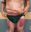
Final post-operative result, demonstrating inset of pedicled right ALT flap to right lower quadrant full thickness abdominal wall defect. Donor site closed with split-thickness skin graft. ALT, anterolateral thigh.
Figure 10
Figure 10
Perianal defect after resection of dermatofibrosarcoma protuberans (DFSP) with exposed rectum.
Figure 11
Figure 11
Final post-operative result of perianal DFSP defect closed with bilateral V-Y gluteal flaps. DFSP, dermatofibrosarcoma protuberans.
Figure 12
Figure 12
Locoregional flap considerations based on anatomic location of defect. TFL, tensor fascia lata; ALT, anterolateral thigh; SGAP, superior gluteal artery perforator; IGAP, inferior gluteal artery perforator; DIEP, deep inferior epigastric perforator.

References

    1. Husain N, Verma N. Current concepts in pathology of soft tissue sarcoma. Indian J Surg Oncol 2011;2:302-8. 10.1007/s13193-012-0134-6 - DOI - PMC - PubMed
    1. Stojadinovic A, Hoos A, Karpoff HM, et al. Soft tissue tumors of the abdominal wall: analysis of disease patterns and treatment. Arch Surg 2001;136:70-9. 10.1001/archsurg.136.1.70 - DOI - PubMed
    1. von Mehren M, Randall RL, Benjamin RS, et al. Soft Tissue Sarcoma, Version 2.2018, NCCN Clinical Practice Guidelines in Oncology. J Natl Compr Canc Netw 2018;16:536-63. 10.6004/jnccn.2018.0025 - DOI - PubMed
    1. Mullinax JE, Zager JS, Gonzalez RJ. Current diagnosis and management of retroperitoneal sarcoma. Cancer Control 2011;18:177-87. 10.1177/107327481101800305 - DOI - PubMed
    1. Shiu MH, Weinstein L, Hajdu SI, et al. Malignant soft-tissue tumors of the anterior abdominal wall. Am J Surg 1989;158:446-51. 10.1016/0002-9610(89)90284-5 - DOI - PubMed