

Macular pigment density changes in central serous chorioretinopathy
- PMID: 30505992
- PMCID: PMC6256073
Macular pigment density changes in central serous chorioretinopathy
Abstract
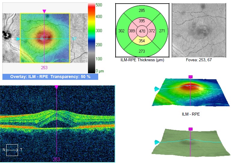
Aim: To present a series of 2 cases of central serous chorioretinopathy and the changes in the macular pigment optical density during the evolution of the disease. Material and methods: A 32-year-old patient presented himself for blurred vision on his LE. The SD OCT imaging revealed serous macular detachment of the neurosensory retina on the LE. The MPOD results were 0.72 on RE and 0.91 on LE. After treatment and resorption of the subretinal fluid, the MPOD values were 0.72 on the RE and 0.82 on the LE. The second patient was a 36-year-old male with metamorphopsia on LE and serous macular detachment on this eye. The MPOD results were 0.43 on RE and 0.58 on the LE and, after treatment, they were 0.38 on the RE and 0.43 on the LE. Conclusions: Central serous chorioretinopathy is a disease of unknown pathophysiology in which we observed a higher MPOD on the eye with CSC than on the fellow eye and a decrease in the MPOD value after the resorption of the subretinal fluid. Abbreviations: L = lutein, Z = zeaxantin, MZ = mezozeaxantin, AMD = age related macular degeneration, MPOD = macular pigment optical density, MP = macular pigment, HFP = Heterochromatic Flicker Photometry, CSC = central serous chorioretinopathy, RE = right eye, LE = left eye.
Keywords: central serous chorioretinopathy; macular pigment optical density.
Figures


Similar articles
-
Central serous chorioretinopathy produces macular pigment profile changes.Optom Vis Sci. 2013 Jul;90(7):e206-12. doi: 10.1097/OPX.0b013e318299386e. Optom Vis Sci. 2013. PMID: 23770658
-
Correlations between internal and external ocular factors and macular pigment optical density.Rom J Ophthalmol. 2018 Jan-Mar;62(1):42-47. Rom J Ophthalmol. 2018. PMID: 29796433 Free PMC article.
-
Macular pigment optical density in central serous chorioretinopathy.Invest Ophthalmol Vis Sci. 2010 Oct;51(10):5219-25. doi: 10.1167/iovs.09-4881. Epub 2010 May 5. Invest Ophthalmol Vis Sci. 2010. PMID: 20445108
-
[Pathophysiology of macular diseases--morphology and function].Nippon Ganka Gakkai Zasshi. 2011 Mar;115(3):238-74; discussion 275. Nippon Ganka Gakkai Zasshi. 2011. PMID: 21476310 Review. Japanese.
-
The role of macular pigment assessment in clinical practice: a review.Clin Exp Optom. 2010 Sep;93(5):300-8. doi: 10.1111/j.1444-0938.2010.00499.x. Epub 2010 Jul 14. Clin Exp Optom. 2010. PMID: 20629669 Review.
Cited by
-
Macular pigment optical density in central serous chorioretinopathy.Ther Adv Ophthalmol. 2021 Mar 3;13:2515841421997195. doi: 10.1177/2515841421997195. eCollection 2021 Jan-Dec. Ther Adv Ophthalmol. 2021. PMID: 33738428 Free PMC article.
References
-
- Ophthalmology AAO . Fundamentals and Principals of Ophthalmology - Retina and Vitreous. San Francisco, CA: 2011.
-
- Alejandra Daruicha AM, Dirania A, Bousquet E, Zhao M, Farmand N, Jaisser F, Behar-Cohen F. Central serous chorioretinopathy: Recent findings and new physiopathology hypothesis. 2015;(48):82–118. - PubMed
-
- Yanoff D. Ophthalmology Part 6 – Retina and vitreous, Section 3 – Anciliary tests, Chapter 6.8 - Fluorescein Angiography, Indocyanine Green Angiography, and Optical Coherence Tomography. 3rd ed. Mosby; 2008.
-
- Bouzas PKEA, Pournaras CJ. Central serous chorioretinopathy and glucocorticoids. Surv Ophthalmol. 2002;(47):431–448. - PubMed
-
- Estera Igras JL, Ratzlaff M, O’Caoimh R, O’Brien C. Evidence of lower macular pigment optical density in chronic open angle glaucoma. Br J Ophthalmol. 2013;(97):994–998. - PubMed
MeSH terms
Substances
LinkOut - more resources
Full Text Sources