. 2018 Sep;2(9):963-971.
doi: 10.1016/j.oret.2018.02.001. Epub 2018 Mar 21.

Spectral-Domain OCT Findings of Retinal Vascular-Avascular Junction in Infants with Retinopathy of Prematurity

Affiliations

Spectral-Domain OCT Findings of Retinal Vascular-Avascular Junction in Infants with Retinopathy of Prematurity

Xi Chen et al. Ophthalmol Retina. 2018 Sep.

Abstract

Purpose: Bedside examination of premature infants at risk for retinopathy of prematurity (ROP) is predominantly performed with ophthalmoscopic en face viewing of the retina. While postmortem retinal microstructures have been studied at the vascular-avascular junction, a critical location for pathogenesis of ROP, to date this has not been possible in vivo. Here we present bedside, non-sedated in vivo cross-sectional imaging and analysis of retinal microstructures at the vascular-avascular junction in infants with ROP using handheld spectral-domain optical coherence tomography (SDOCT).

Design: Prospective observational study.

Participants: Eleven preterm infants consented for research imaging during ROP screening examinations.

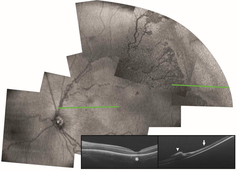
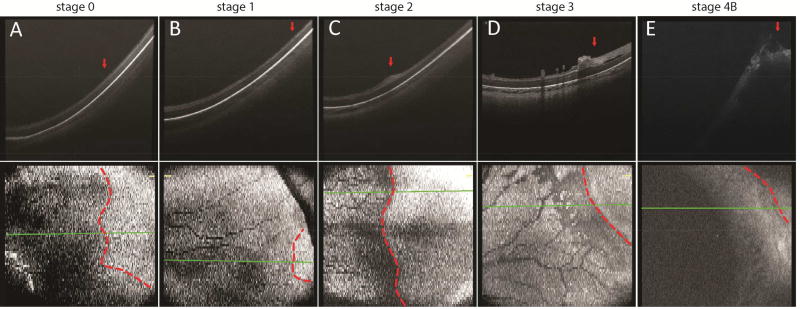
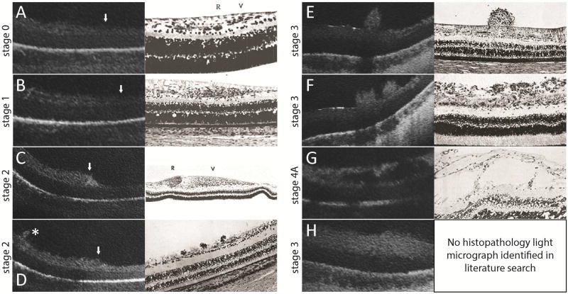
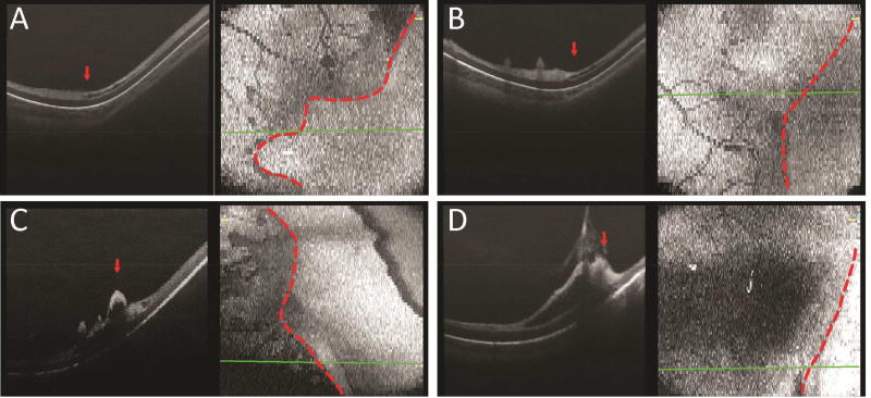
Methods: We imaged the vascular-avascular junction in the temporal retina using a SDOCT system (Envisu, Bioptigen Inc., NC) in 18 eyes from 11 preterm infants with zone I or II, stage 0 through 4 ROP. B-scan and en face images were analyzed and compared to historical light micrographs.

Main outcome measures: SDOCT morphology at the vascular-avascular junction.

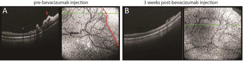
Results: Multiple bedside SDOCT findings at the vascular-avascular junction were comparable to historic light micrographs: thickened inner retinal ridge structure in stage 2 ROP was comparable to thickened vanguard and rear guard cells in micrographs; vascular tufts on the posterior retinal surface in stage 2 ROP, broad arcs of neovascularization above the retina in stage 3 ROP, and splitting of inner retinal layers into clefts on either side of neovascularization mimicked findings of historic light micrographs. A unique findings was thickening of the avascular inner retinal band adjacent to neovascularization. On SDOCT imaging over several weeks, neovascularization and retinal clefts diminished after intravitreal bevacizumab therapy.

Conclusions: Retinal morphology at the vascular-avascular junction imaged with handheld SDOCT is consistent with known histopathology, and provide the advantage of monitoring change in vivo over time. These unique findings provide new insights into preterm retinal neurovascular development in ROP.


Conflict of interest statement

Conflict of Interest: No other authors have financial disclosures. No authors have a proprietary interest in the current study.

Figures

Figure 1
Figure 1
En face view and representative OCT B-scan of posterior pole and temporal retina in a preterm infant with zone II stage 3 ROP at the time of ROP screening examination. B-scan at the macular area (green line, left) showed absence of subfoveal photoreceptor ellipsoid zone (left panel, star). B-scan at the temporal retinal periphery (green line, right) showed preretinal neovascular tissue (right panel, arrowhead), inner retinal split, and inner retinal thickening (right panel, arrow).
Figure 2
Figure 2
Representative OCT B-scan and en face images across the vascular-avascular junction in the temporal peripheral retina in preterm infants with zone I stage 0 (A), zone II stage 1 (B), zone I stage 2 (C), zone I stage 3 (D), and zone I stage 4B (E) ROP. The vascular-avascular junction was delineated on the en face retina view (bottom panel, dotted line) and its respective position marked on the B-scan image (top panel, arrow). The retinal and choroidal thickness at the vascular-avascular junction on these B-scans, as well as retinal thickness 1000 µm anterior or posterior to the vascular-avascular junction are shown in Supplementary Table 1.
Figure 3
Figure 3
SDOCT images (left) are correlated with historic representative light micrographs (right) at the vascular-avascular junction (A–D, white arrowhead with vascularized retina on the left side of the images) or adjacent to the vascular-avascular junction (E–G, posterior, and H, anterior) in eyes with ROP. A: Stage 0 ROP: in vascularized retina a faint hyporeflective band divided the inner retinal hyperreflective layer which gradually tapered to a single hyperreflective band in avascular retina. This was comparable to the normal angiogenesis pattern reported by Foos (adapted image), although the retinal layers were not as evident on OCT imaging compared to histopathology studies. B: Stage 1 ROP: a similar three-layered inner retina on the left gradually tapered to a one-layered structure in avascular retina. Similar to Foos, the total thickness of the vascularized retina appeared to be greater than in stage 0 ROP. (also see Supplementary Table 1 thicknesses) C: Stage 2 ROP: pronounced thickening at the vascular-avascular junction was consistent with the ridge structure with hypertrophied anterior vanguard and posterior rear guard cells reported by Foos. As with all OCT imaging, specific cellular elements could not be distinguished. D: Small neovascular buds, not noticed on clinical examination, could be seen on OCT in stage 2 ROP (asterisk). This was similar to the small elevations on the retinal surface observed by Reese, which were presumed to be early endothelial proliferation. E, F: Stage 3 ROP: neovascular buds (E) and bands (F) frequented the preretinal surface. Their configurations were also consistent with neovascular findings reported by Reese. G: Stage 4A ROP: there were schisis-like changes in the inner retinal layers posterior to the presumed area of retinal detachment. These schisis-like changes were often cavitary, and appeared similar to the large cystic spaces in the nerve fiber layer in later stages of proliferative disease reported by Kushner. H: In stage 3 or 4 ROP, there was often diffuse thickening of the inner retinal layer of avascular retina. No correlating histopathology light micrograph was identified in our literature search.
Figure 4
Figure 4
Representative OCT B-scan and en face images of the temporal retina in preterm infants with stage 3 ROP, with an inner retinal split, a hyporeflective lucency within the inner hyperreflective band, anterior to (A), at (B), and posterior to (C) the vascular-avascular junction. Focal vitreoretinal traction was also observed in some eyes with stage 3 ROP (D).
Figure 5
Figure 5
Representative OCT B-scan and en face images of the temporal retina in a preterm infant with zone I stage 3 ROP, prior to (A) and three weeks after (B) intravitreal bevacizumab injection, showing regression of preretinal neovascular elevations and further vascularization of the previously avascular retina compared to images in (A).
Figure 6
Figure 6
Schematic model of retinal development at the vascular-avascular junction on OCT imaging during different stages of ROP. The dash lines represent retinal layers observed on OCT: white dash line corresponds to outer plexiform layer and grey dash line represents the inner retinal hyperreflective band.

References

    1. Palmer EA, Flynn JT, Hardy RJ, et al. Incidence and early course of retinopathy of prematurity. The Cryotherapy for Retinopathy of Prematurity Cooperative Group. Ophthalmology. 1991;98(11):1628–40. - PubMed
    1. Quinn GE, Dobson V, Barr CC, et al. Visual acuity in infants after vitrectomy for severe retinopathy of prematurity. Ophthalmology. 1991;98(1):5–13. - PubMed
    1. Tasman W. Retinopathy of prematurity: do we still have a problem?: the Charles L. Schepens lecture. Arch Ophthalmol. 2011;129(8):1083–6. - PubMed
    1. Hartnett ME. Vascular endothelial growth factor antagonist therapy for retinopathy of prematurity. Clin Perinatol. 2014;41(4):925–43. - PMC - PubMed
    1. Mintz-Hittner HA, Kennedy KA, Chuang AZ, Group B-RC. Efficacy of intravitreal bevacizumab for stage 3+ retinopathy of prematurity. N Engl J Med. 2011;364(7):603–15. - PMC - PubMed