. 2018 Nov 30;29(12):181.
doi: 10.1007/s10856-018-6190-x.

Bio-inspired melanin nanoparticles induce cancer cell death by iron adsorption

Affiliations

Bio-inspired melanin nanoparticles induce cancer cell death by iron adsorption

James Perring et al. J Mater Sci Mater Med. .

Abstract

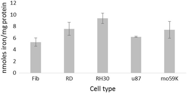
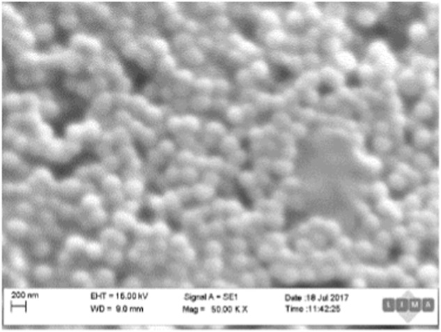
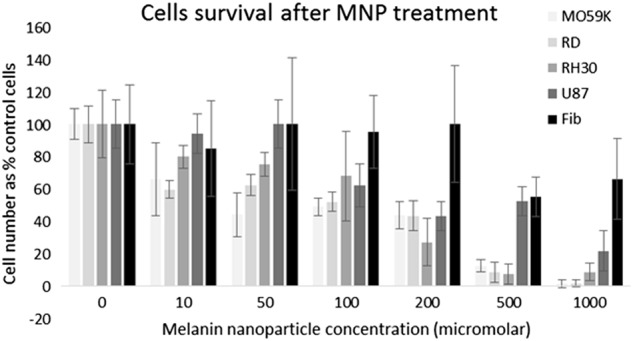
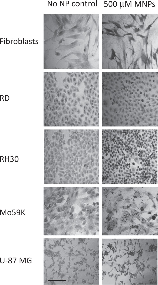
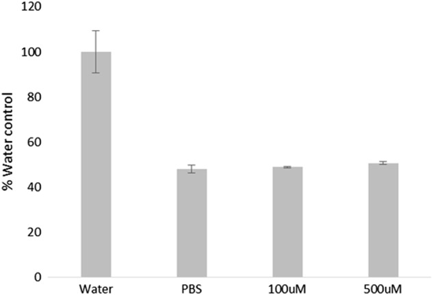
Dysregulation of iron metabolism is a common characteristic of cancer cells. The rapid proliferation of the tumour cells means that there is an increased dependence upon iron compared to healthy cells. Chelation of iron can be undertaken with a number of different compounds, however, simply lowering systemic iron levels to control tumour growth is not possible since iron is essential for cellular metabolism in the rest of the body. Nanoparticulate iron chelators could overcome this difficulty by targeting to the tumour either by the passive enhanced permeation and retention effect, or by targeting ligands on the surface. Nanoparticles were prepared from melanin, which is a naturally occurring pigment that is widely distributed within the body, but that can chelate iron. The prepared nanoparticles were shown to be ~220 nm, and could adsorb 16.45 mmoles iron/g melanin. The nanoparticles showed no affect on control fibroblast cells at a concentration of 200 μM, whereas the immortalised cancer cell lines showed at least 56% reduction in cell growth. At a concentration of 1 mM melanin nanoparticles the cell growth could be reduced by 99% compared to the control. The nanoparticles also show no significant haemotoxicity, even at concentration of 500 μM. Melanin nanoparticles are therefore a viable prospect for destroying cancer cells via iron starvation.


Conflict of interest statement

The authors declare that they have no conflict of interest.

Figures

Fig. 1
Fig. 1
Iron levels in RMS and GBM cell lines. Cells from the different origins were homogenised and total iron measured relative to the protein concentration. Data shown represents n = 3 sample repeats, and n = 3 independent experiments. Data is shown as average ± standard deviation
Fig. 2
Fig. 2
Effects of varying concentrations of desferrioxamine (DFO) on cell survival. Cell lines were incubated with DFO for 24 h to assess the effect on cell survival. (●) Fibroblast cells (■) RH30 cells (▲) RD cells (♦) U-87 MG (x) Fibroblasts. Data shown represents n = 3 sample repeats, and n = 2 independent experiments. Data is shown as average ± standard deviation
Fig. 3
Fig. 3
Scanning Electron Microscope image of melanin nanoparticles. The image shows spherical melanin nanoparticles. The scale bar is 200 nm
Fig. 4
Fig. 4
Melanin nanoparticle Iron chelation. Melanin nanoparticles (1 mM and 5 mM) were incubated with an iron sulphate solution (1, 2, 6.8 and 8 mM). The particles were incubated in the iron solutions for either 24 or 48 h. Data shown represents n = 3 sample repeats, and n = 2 independent repeats. Data shown is average ±standard deviation
Fig. 5
Fig. 5
Cell survival after iron chelation. Cell lines were incubated with increasing concentrations of melanin nanoparticles, for 24 h. Data shown represents n = 3 sample repeats, and n = 3 independent repeats. Data shown is average ± standard deviation
Fig. 6
Fig. 6
Iron staining of immortalised cells. Cells were incubated with melanin nanoparticles for 24 h and compared to controls. The cells were fixed and stained with Perl’s reagent and Neutral Red. Representative images are shown. Scale bar shows 100 µm
Fig. 7
Fig. 7
Haemotoxicity assay of melanin nanoparticles. Blood was incubated with either water, PBS, or melanin nanoparticles, for 24 h. The serum was analysed spectroscopically to detect whether blood cell lysis had occurred. Data shown represents n = 3 sample repeats, and n = 3 independent repeats. Data shown is average ± standard deviation

References

    1. Manz D, Blanchette N, Paul B, Torti F, Torti S. Iron and cancer: recent insights. Ann N Y Acad Sci. 2016;1368:149–61. doi: 10.1111/nyas.13008. - DOI - PMC - PubMed
    1. Torti S, Torti F. Iron and cancer: more ore to be mined. Nat Rev Cancer. 2013;13:342–55. doi: 10.1038/nrc3495. - DOI - PMC - PubMed
    1. Daniels T, Bernabeu E, Rodríguez J, et al. The transferrin receptor and the targeted delivery of therapeutic agents against cancer. Biochim Biophys Acta. 2012;1820:291–317. doi: 10.1016/j.bbagen.2011.07.016. - DOI - PMC - PubMed
    1. Radulescu S, Brookes M, Salgueiro P, et al. Luminal iron levels govern intestinal tumorigenesis after Apc loss in vivo. Cell Rep. 2012;2:270–82. doi: 10.1016/j.celrep.2012.07.003. - DOI - PubMed
    1. Pinnix Z, Miller L, Wang W, et al. Ferroportin and iron regulation in breast cancer progression and prognosis. Sci Transl Med. 2010;2:43–56. doi: 10.1126/scitranslmed.3001127. - DOI - PMC - PubMed

LinkOut - more resources