. 2019 Feb;97(2):189-199.
doi: 10.1007/s00109-018-1719-5. Epub 2018 Nov 30.

Effect of DJ-1 on the neuroprotection of astrocytes subjected to cerebral ischemia/reperfusion injury

Affiliations

Effect of DJ-1 on the neuroprotection of astrocytes subjected to cerebral ischemia/reperfusion injury

Li Peng et al. J Mol Med (Berl). 2019 Feb.

Abstract

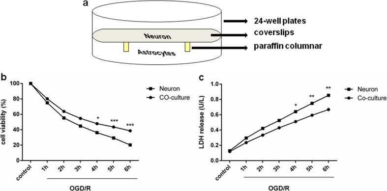
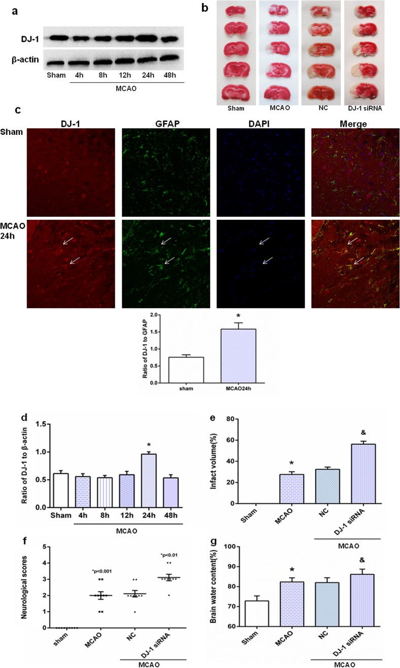
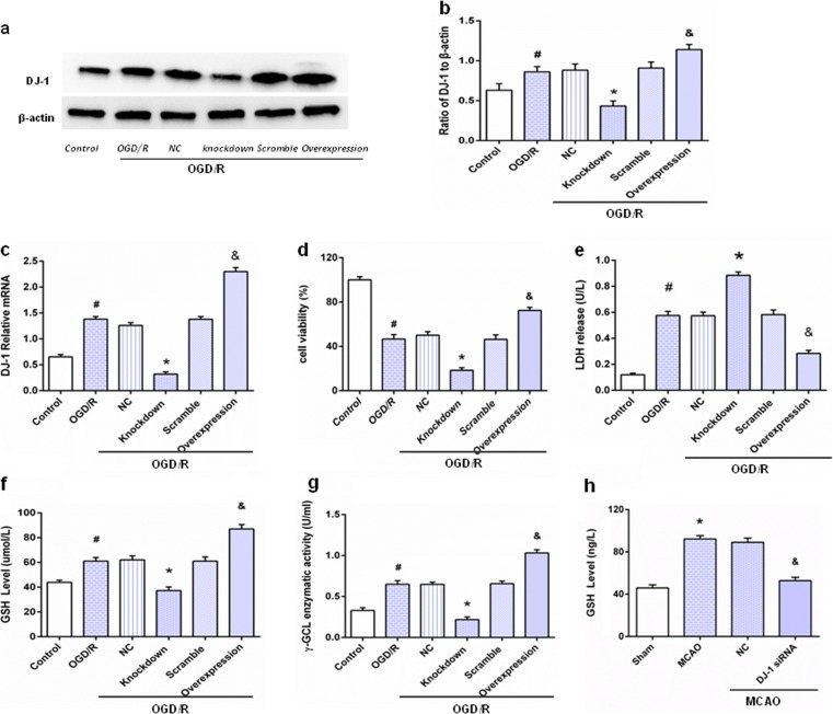
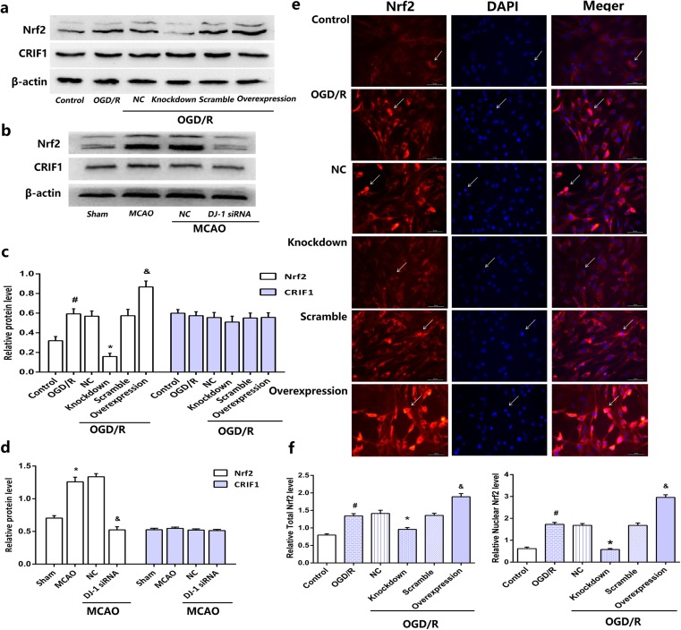
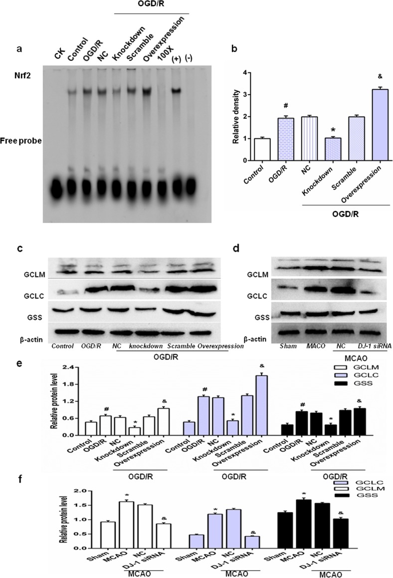
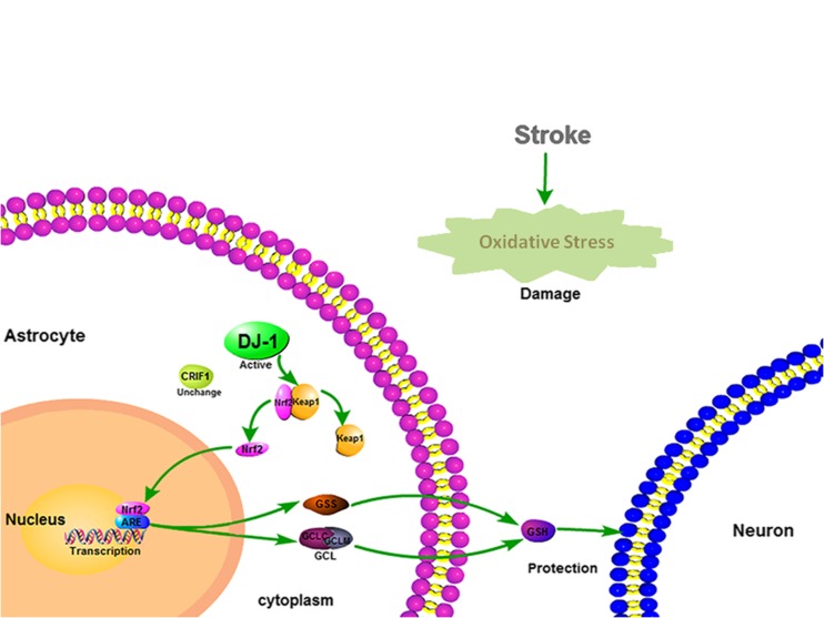
Astrocytes are involved in neuroprotection, and DJ-1 is an important antioxidant protein that is abundantly expressed in reactive astrocytes. However, the role of DJ-1 in astrocytes' neuroprotection in cerebral ischemia/reperfusion injury and its potential mechanism is unclear. Thus, to explore effects and mechanisms of DJ-1 on the neuroprotection of astrocytes, we used primary co-cultures of neurons and astrocytes under oxygen and glucose deprivation/reoxygenation in vitro and transient middle cerebral artery occlusion/reperfusion in vivo to mimic ischemic reperfusion insult. Lentiviral was used to inhibit and upregulate DJ-1 expression in astrocytes, and DJ-1 siRNA blocked DJ-1 expression in rats. Inhibiting DJ-1 expression led to decreases in neuronal viability. DJ-1 knockdown also attenuated total and nuclear Nrf2 and glutathione (GSH) levels in vitro and vivo. Similarly, loss of DJ-1 decreased Nrf2/ARE-binding activity and expression of Nrf2/ARE pathway-driven genes. Overexpression of DJ-1 yielded opposite results. This suggests that the mechanism of action of DJ-1 in astrocyte-mediated neuroprotection may involve regulation of the Nrf2/ARE pathway to increase GSH after cerebral ischemia/reperfusion injury. Thus, DJ-1 may be a new therapeutic target for treating ischemia/reperfusion injury. KEY MESSAGES: Astrocytes protect neurons in co-culture after OGD/R DJ-1 is upregulated in astrocytes and plays an important physiological roles in neuronal protection under ischemic conditions DJ-1 protects neuron by the Nrf2/ARE pathway which upregulates GSH.

Keywords: Astrocyte; Co-culture; DJ-1; GSH; I/R injury; Nrf2/ARE.


Conflict of interest statement

Conflict of interest

The authors declare that they have no conflicts of interest.

Figures

Fig. 1
Fig. 1
Co-culture with astrocytes increased neuronal survival after OGD/R. a Non-contact neuron-astrocyte co-culture system. b Cell viability data. n = 6 of samples/group from an experiment, three independent experiments were carried out. c LDH release. n = 6 of samples/group from an experiment, three independent experiments were carried out. OGD/R = oxygen-glucose deprivation/reoxygenation. Values are expressed as mean ± SEM. Neuronal monoculture vs. co-culture; *p < 0.05, **p < 0.01, ***p < 0.001
Fig. 2
Fig. 2
DJ-1 ameliorated cerebral (I/R) injury after MCAO/R. a Western blot of DJ-1. d Ratios of DJ-1 relative to β-actin. In the 24-h group, DJ-1 levels increased compared with the sham group. Values are mean ± SEM. *p < 0.05. n = 3 of samples/group from an experiment, three independent experiments were carried out. c Immunohistochemistry for DJ-1 and semi-quantitation to determine percentage of DJ-1-postive cells relative to GFAP-positive cells in astrocytes. In the 24-h MCAO group, DJ-1 was expressed at higher levels in reactive astrocytes in cortical infarct regions compared with controls. Laser scanning confocal microscope was used to assess expression. DJ-1 expression in astrocytes is indicated by red fluorescence. GFAP expression in astrocytes is indicated by green fluorescence. Cell nuclei were stained with DAPI. Original magnification, 400×. Values are expressed as mean ± SEM. *p < 0.05 vs. sham. n = 3 of samples/group from an experiment, three independent experiments were carried out. b, e Infarct volume of brain. f Neurological scores. g Brain water content. Infarct volume, neurological scores, brain water content were significantly increased in the DJ-1 siRNA group compared with the MCAO and NC groups. Values are mean ± SEM. *p < 0.01 vs. sham; &p < 0.05 vs. MCAO, n = 3 of samples/group from an experiment, three independent experiments were carried out
Fig. 3
Fig. 3
DJ-1 expression in astrocytes varied with OGD duration. After OGD/R, astrocytes were harvested. a Western blot analysis of DJ-1. b Ratios of DJ-1 relative to β-actin showing that DJ-1 in the 5-h group increased compared with controls. c q-PCR data was consistent with results of Western blots. d Immunocytochemistry to measure DJ-1 expression in astrocytes. Fluorescence microscopy was used to assess expression. DJ-1 expression in astrocytes is indicated by red fluorescence. GFAP expression in astrocytes is indicated by green fluorescence. Cell nuclei were stained with DAPI. Original magnification, 200×. e Semi-quantitation to determine percentage of DJ-1-postive cells relative to GFAP-positive cells in astrocytes. Values are mean ± SEM. *p < 0.05, n = 3 of samples/group from an experiment, three independent experiments were carried out
Fig. 4
Fig. 4
Astrocytic DJ-1 protected neurons by upregulating GSH levels. After OGD for 5 h followed by reoxygenation for 24 h, astrocytes, neurons, and culture medium were harvested. a Western blot of DJ-1. b Ratios of DJ-1 relative to β-actin. DJ-1 expression was reduced in the knockdown group and upregulated in the overexpression group. c q-PCR results were consistent with Western blots. Values are expressed as mean ± SEM. #p < 0.05 vs. control; *p < 0.01 vs. OGD/R; &p < 0.01 vs. OGD/R. n = 3 of samples/group from an experiment, three independent experiments were carried out. d Neuronal viability after exposure to OGD/R. e LDH in the culture medium. f GSH in the culture medium. g γ-GCL in the culture medium. h GSH in rats. Values represent mean ± SEM. #p < 0.05 vs. control; *p < 0.01 vs. OGD/R; &p < 0.05 vs. OGD/R. n = 6 of samples/group from an experiment, three independent experiments were carried out. Values are mean ± SEM. *p < 0.01 vs. sham; &p < 0.01 vs. MCAO, n = 3 of samples/group from an experiment, three independent experiments were carried out
Fig. 5
Fig. 5
Effects of DJ-1 on expression of Nrf2. a After OGD for 5 h followed by 24 h of reoxygenation, astrocytes were harvested. Western blot for Nrf2 and CRIF1 in astrocytes. b After 24 h of reperfusion, brains were collected. Western blot for Nrf2 and CRIF1 in rats. c, d Ratios of Nrf2 and CRIF1 relative to β-actin in astrocytes and in rats, respectively. e Immunocytochemistry assays to measure total Nrf2 and nuclear Nrf2 in astrocytes. Fluorescence microscopy was used to assess expression. Nrf2 expression in astrocytes is indicated by red fluorescence. Cell nuclei were stained with DAPI. Original magnification, 200×. f Semi-quantitation to determine total number of Nrf2-positive cells and number of nuclear Nrf2-positive cells in astrocytes. Values are mean ± SEM. #p < 0.05 vs. control; *p < 0.05 vs. OGD/R; and &p < 0.05 vs. OGD/R. n = 3 of samples/group from an experiment, three independent experiments were carried out. Values are mean ± SEM. *p < 0.01 vs. sham; &p < 0.01 vs. MCAO, n = 3 of samples/group from an experiment, three independent experiments were carried out
Fig. 6
Fig. 6
DJ-1 regulates Nrf2/ARE binding activity and Nrf2/ARE-driven gene expression. After OGD for 5 h followed by 24 h of reoxygenation, astrocytes were harvested. And after 24 h of reperfusion, brains were collected. a EMSA analysis of Nrf2/ARE binding. b Semiquantitative analysis of Nrf2/ARE binding. CK, 100x, (+) and (−) indicate controls. c Western blot for GCLM, GCLC, and GSS in astrocytes. d Western blot for GCLM, GCLC, and GSS in rats. e, f Ratios of GCLM, GCLC, and GSS relative to β-actin in astrocytes and in rats, respectively. Values are mean ± SEM. #p < 0.01 vs. control; *p < 0.01 vs. OGD/R; and p < 0.05 vs. OGD/R. n = 3 of samples/group from an experiment, three independent experiments were carried out. Values are mean ± SEM. *p < 0.05 vs. sham; &p < 0.01 vs. MCAO, n = 3 of samples/group from an experiment, three independent experiments were carried out
Fig. 7
Fig. 7
Mechanisms of action of DJ-1 in astrocytes. After oxidative stress induced by cerebral ischemia and reperfusion, DJ-1 is expressed in immunoreactive astrocytes. DJ-1 facilitates Nrf2 translocation to the nucleus by preventing binding with Keap1. Nrf2 binds to ARE and upregulates expression of GCLC, GCLM, and GSS, which regulate synthesis of GSH

References

    1. Juurlink BH, Sweeney MI. Mechanisms that result in damage during and following cerebral ischemia. Neurosci Biobehav Rev. 1997;21(2):121–128. doi: 10.1016/S0149-7634(96)00001-2. - DOI - PubMed
    1. Barreto G, E. White R, Ouyang Y, Xu L, G. Giffard R. Astrocytes: targets for neuroprotection in stroke. Cent Nerv Syst Agents Med Chem. 2011;11(2):164–173. doi: 10.2174/187152411796011303. - DOI - PMC - PubMed
    1. Heneka MT, Rodriguez JJ, Verkhratsky A. Neuroglia in neurodegeneration. Brain Res Rev. 2010;63(1–2):189–211. doi: 10.1016/j.brainresrev.2009.11.004. - DOI - PubMed
    1. Park LC, Zhang H, Gibson GE. Co-culture with astrocytes or microglia protects metabolically impaired neurons. Mech Ageing Dev. 2001;123(1):21–27. doi: 10.1016/S0047-6374(01)00336-0. - DOI - PubMed
    1. Wang XF, Cynader MS. Effects of astrocytes on neuronal attachment and survival shown in a serum-free co-culture system. Brain Res Brain Res Protoc. 1999;4(2):209–216. doi: 10.1016/S1385-299X(99)00019-7. - DOI - PubMed

Publication types

LinkOut - more resources