

Repeatability of 18 F-FDG PET radiomic features: A phantom study to explore sensitivity to image reconstruction settings, noise, and delineation method
- PMID: 30506687
- PMCID: PMC7380016
- DOI: 10.1002/mp.13322
Repeatability of 18 F-FDG PET radiomic features: A phantom study to explore sensitivity to image reconstruction settings, noise, and delineation method
Abstract
Background: 18 F-fluoro-2-deoxy-D-Glucose positron emission tomography (18 F-FDG PET) radiomics has the potential to guide the clinical decision making in cancer patients, but validation is required before radiomics can be implemented in the clinical setting. The aim of this study was to explore how feature space reduction and repeatability of 18 F-FDG PET radiomic features are affected by various sources of variation such as underlying data (e.g., object size and uptake), image reconstruction methods and settings, noise, discretization method, and delineation method.
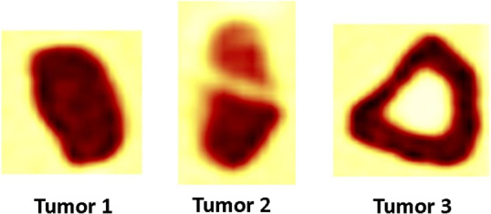
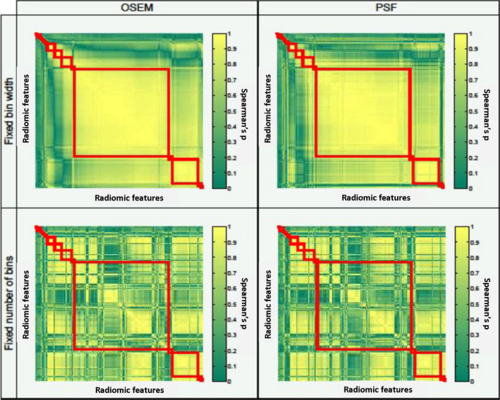
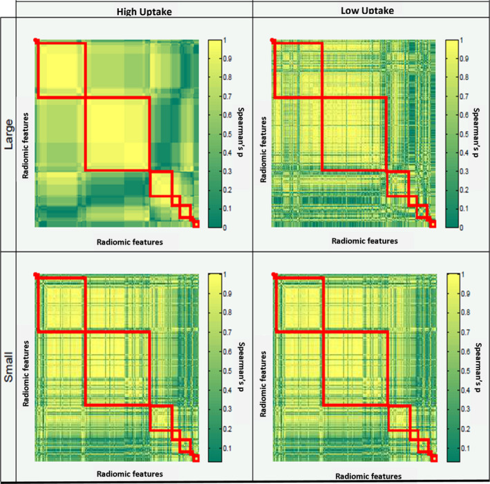
Methods: The NEMA image quality phantom was scanned with various sphere-to-background ratios (SBR), simulating different activity uptakes, including spheres with low uptake, that is, SBR smaller than 1. Furthermore, images of a phantom containing 3D printed inserts reflecting realistic heterogeneity uptake patterns were acquired. Data were reconstructed using various matrix sizes, reconstruction algorithms, and scan durations (noise). For every specific reconstruction and noise level, ten statistically equal replicates were generated. The phantom inserts were delineated using CT and PET-based segmentation methods. A total of 246 radiomic features was extracted from each image dataset. Images were discretized with a fixed number of 64 bins (FBN) and a fixed bin width (FBW) of 0.25 for the high and a FBW of 0.05 for the low uptake data. In terms of feature reduction, we determined the impact of these factors on the composition of feature clusters, which were defined on the basis of Spearman's correlation matrices. To assess feature repeatability, the intraclass correlation coefficient was calculated over the ten replicates.
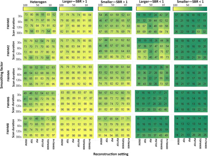
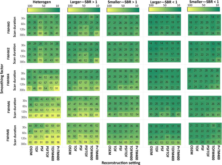
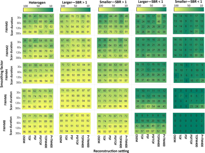
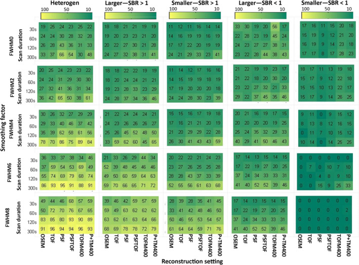
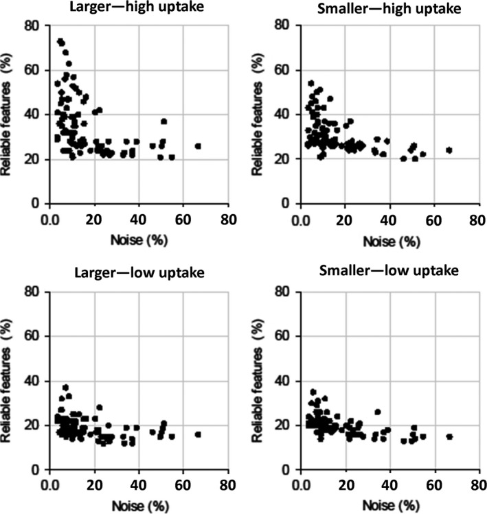
Results: In general, larger spheres with high uptake resulted in better repeatability compared to smaller low uptake spheres. In terms of repeatability, features extracted from heterogeneous phantom inserts were comparable to features extracted from bigger high uptake spheres. For example, for an EARL-compliant reconstruction, larger and smaller high uptake spheres yielded good repeatability for 32% and 30% of the features, while the heterogeneous inserts resulted in 34% repeatable features. For the low uptake spheres, this was the case for 22% and 20% of the features for bigger and smaller spheres, respectively. Images reconstructed with point-spread-function (PSF) resulted in the highest repeatability when compared with OSEM or time-of-flight, for example, 53%, 30%, and 32% of repeatable features, respectively (for unsmoothed data, discretized with FBN, 300 s scan duration). Reducing image noise (increasing scan duration and smoothing) and using CT-based segmentation for the low uptake spheres yielded improved repeatability. FBW discretization resulted in higher repeatability than FBN discretization, for example, 89% and 35% of the features, respectively (for the EARL-compliant reconstruction and larger high uptake spheres).
Conclusion: Feature space reduction and repeatability of 18 F-FDG PET radiomic features depended on all studied factors. The high sensitivity of PET radiomic features to image quality suggests that a high level of image acquisition and preprocessing standardization is required to be used as clinical imaging biomarker.
Keywords: 18F-FDG PET/CT radiomic features; delineation; image reconstruction settings.
© 2018 The Authors. Medical Physics published by Wiley Periodicals, Inc. on behalf of American Association of Physicists in Medicine.
Conflict of interest statement
The authors have no relevant conflicts of interest to disclose.
Figures
Similar articles
-
Experimental Multicenter and Multivendor Evaluation of the Performance of PET Radiomic Features Using 3-Dimensionally Printed Phantom Inserts.J Nucl Med. 2020 Mar;61(3):469-476. doi: 10.2967/jnumed.119.229724. Epub 2019 Aug 16. J Nucl Med. 2020. PMID: 31420497 Free PMC article.
-
Effects of Tracer Uptake Time in Non-Small Cell Lung Cancer 18F-FDG PET Radiomics.J Nucl Med. 2022 Jun;63(6):919-924. doi: 10.2967/jnumed.121.262660. Epub 2021 Dec 21. J Nucl Med. 2022. PMID: 34933890 Free PMC article.
-
Reproducibility of F18-FDG PET radiomic features for different cervical tumor segmentation methods, gray-level discretization, and reconstruction algorithms.J Appl Clin Med Phys. 2017 Nov;18(6):32-48. doi: 10.1002/acm2.12170. Epub 2017 Sep 11. J Appl Clin Med Phys. 2017. PMID: 28891217 Free PMC article.
-
A Role for FDG PET Radiomics in Personalized Medicine?Semin Nucl Med. 2020 Nov;50(6):532-540. doi: 10.1053/j.semnuclmed.2020.05.002. Epub 2020 Jun 15. Semin Nucl Med. 2020. PMID: 33059822 Free PMC article. Review.
-
Impact of Preprocessing Parameters in Medical Imaging-Based Radiomic Studies: A Systematic Review.Cancers (Basel). 2024 Jul 26;16(15):2668. doi: 10.3390/cancers16152668. Cancers (Basel). 2024. PMID: 39123396 Free PMC article. Review.
Cited by
-
Joint EANM/SNMMI guideline on radiomics in nuclear medicine : Jointly supported by the EANM Physics Committee and the SNMMI Physics, Instrumentation and Data Sciences Council.Eur J Nucl Med Mol Imaging. 2023 Jan;50(2):352-375. doi: 10.1007/s00259-022-06001-6. Epub 2022 Nov 3. Eur J Nucl Med Mol Imaging. 2023. PMID: 36326868 Free PMC article.
-
A heterogeneous phantom study for investigating the stability of PET images radiomic features with varying reconstruction settings.Front Nucl Med. 2023 Feb 14;3:1078536. doi: 10.3389/fnume.2023.1078536. eCollection 2023. Front Nucl Med. 2023. PMID: 39380957 Free PMC article.
-
Physical imaging phantoms for simulation of tumor heterogeneity in PET, CT, and MRI: An overview of existing designs.Med Phys. 2020 Apr;47(4):2023-2037. doi: 10.1002/mp.14045. Epub 2020 Feb 12. Med Phys. 2020. PMID: 31981214 Free PMC article. Review.
-
Clinical use of positron emission tomography for radiotherapy planning - Medical physics considerations.Z Med Phys. 2023 Feb;33(1):13-21. doi: 10.1016/j.zemedi.2022.09.001. Epub 2022 Oct 20. Z Med Phys. 2023. PMID: 36272949 Free PMC article. Review.
-
The application of radiomics in predicting gene mutations in cancer.Eur Radiol. 2022 Jun;32(6):4014-4024. doi: 10.1007/s00330-021-08520-6. Epub 2022 Jan 20. Eur Radiol. 2022. PMID: 35048135 Review.
References
-
- Huang W, Fan M, Liu B, et al. Value of metabolic tumor volume on repeated 18F‐FDG PET/CT for early prediction of survival in locally advanced non‐small cell lung cancer treated with concurrent chemoradiotherapy. J Nucl Med. 2014;55:1584–1590. - PubMed
-
- Hatt M, Majdoub M, Vallieres M, et al. 18F‐FDG PET uptake characterization through texture analysis: investigating the complementary nature of heterogeneity and functional tumor volume in a multi‐cancer site patient cohort. J Nucl Med. 2015;56:38–44. - PubMed
MeSH terms
Substances
Grants and funding
LinkOut - more resources
Full Text Sources
