. 2019 Feb;23(2):1386-1395.
doi: 10.1111/jcmm.14041. Epub 2018 Dec 3.

Induction of Krüppel-like factor 2 reduces K/BxN serum-induced arthritis

Affiliations

Induction of Krüppel-like factor 2 reduces K/BxN serum-induced arthritis

Manjusri Das et al. J Cell Mol Med. 2019 Feb.

Abstract

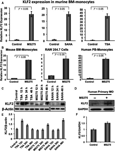
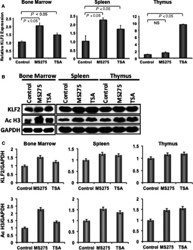
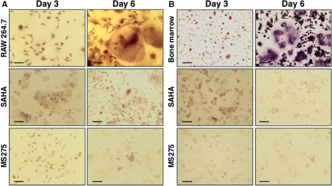
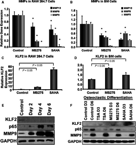
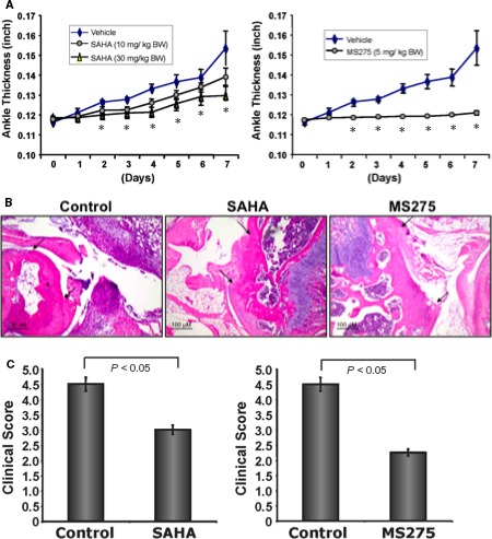
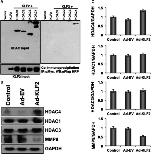
Krüppel-like factor 2 (KLF2) critically regulates activation and function of monocyte, which plays important pathogenic role in progressive joint destruction in rheumatoid arthritis (RA). It is yet to be established the molecular basis of KLF2-mediated regulation of monocytes in RA pathogenesis. Herein, we show that a class of compound, HDAC inhibitors (HDACi) induced KLF2 expression in monocytes both in vitro and in vivo. KLF2 level was also elevated in tissues, such as bone marrow, spleen and thymus in mice after infusion of HDACi. Importantly, HDACi significantly reduced osteoclastic differentiation of monocytes with the up-regulation of KLF2 and concomitant down-regulation of matrixmetalloproteinases both in the expression level as well as in the protein level. In addition, HDACi reduced K/BxN serum-induced arthritic inflammation and joint destruction in mice in a dose-dependent manner. Finally, co-immunoprecipitation and overexpression studies confirmed that KLF2 directly interacts with HDAC4 molecule in cells. These findings provide mechanistic evidence of KLF2-mediated regulation of K/BxN serum-induced arthritic inflammation.

Keywords: HDAC inhibitors; KLF2; inflammation; monocytes; osteoclasts; rheumatoid arthritis.


Figures

Figure 1
Figure 1
HDAC inhibitor induces KLF2 expression in monocytes. (A) Real‐time RTPCR was performed to evaluate the level of relative KLF2 expression in RNA isolated from mouse primary bone marrow‐derived monocytes (n = 3), after treating with 10 nmol/L of various HDACi (MS275, SAHA or TSA) for 24 h, and without stimulus (DMSO) was considered as a base line control of KLF2 expression, and the relative fold expression was graphically presented. GAPDH was considered as an internal control. Experiments were performed in triplicate. (B) Similar experiment was performed with mouse primary bone marrow‐derived monocytes (n = 3), mouse monocyte cell line (RAW 264.7) and human primary peripheral blood‐derived monocytes (n = 3) after treating with 10 nmol/L HDACi (MS275) for 24 h, and data are presented graphically. (C, D) Similar experiments were performed with mouse bone marrow monocytes, RAW 264.7, and human primary peripheral blood‐derived monocytes after stimulation with 10 nmol/L of TSA, or MS275 or SAHA, and proteins were evaluated at various time‐points as stated, and evaluated for KLF2 expression using Western blot methods using β‐actin as an internal control. (E, F) Quantified Western blot data of figures C, D are presented graphically
Figure 2
Figure 2
HDAC inhibitor induces KLF2 expression in various tissues in mice. (A) Three mice per group were injected (ip) with HDACi, and after 24 h bone marrow, spleen and thymus were harvested. Tissues were homogenized and part was subjected to real‐time RTPCR to evaluate the level of relative KLF2 expression in RNA. Vehicle control injected mice was considered as a base line control for KLF2 expression and the relative fold expression was graphically presented for various tissues. GAPDH was considered as an internal control. PCR experiments were performed in triplicate. (B) Other part of the tissues was subjected to protein extraction, and followed by Western blot analysis for KLF2 expression. The levels of KLF2 expression and acetylated Histone 3 were shown along with GAPDH (as an internal control). (C) Quantified Western blot data of stated molecules are presented graphically
Figure 3
Figure 3
HDAC inhibitors reduce osteoclastic differentiation of monocytes. Murine monocytic cell line (RAW 264.7) (A) and murine primary bone marrow cells (B) were subjected to osteoclastic differentiation in the presence or absence of HDACi, such as SAHA (10 nmol/L) and MS275 (10 nmol/L). TRAP staining was performed during the course of osteoclastic differentiation at days 3 and 6. Images were depicted here. Scale bar indicates 10 μmol/L
Figure 4
Figure 4
Reduced osteoclastic differentiation is associated with reduced levels of MMPs and increased level of KLF2. Total RNA was isolated from murine monocytic cell (RAW 264.7) (A, C) and murine primary bone marrow cells (B, D) that were subjected to osteoclastic differentiation in the presence or absence of HDACi, such as SAHA (10 nmol/L), MS275 (10 nmol/L) or TSA (10 nmol/L), and were subjected to real‐time RTPCR analysis for expression levels of MMP3, 9 and 13 along with KLF2 keeping GAPDH as an internal control, shown graphically. RTPCR experiments were performed in triplicate. The experiment was performed at least three times, and (*) indicates statistical significance (P < 0.05) when compared to the respective controls. (E) Western blot analysis was performed during the course of osteoclastic differentiation of RAW 264.7 cells for KLF2, NFκB (p65) and MMP9 proteins keeping GAPDH as an internal control. (F) Western blot analysis was performed during the course of osteoclastic differentiation of RAW 264.7 cells as indicated for KLF2, NFκB (p65) and MMP9 proteins keeping GAPDH as an internal control in the presence or absence of HDACi
Figure 5
Figure 5
HDAC inhibitors reduce K/BxN serum‐induced arthritic inflammation, and bone and cartilage damage in mice. Group of C57BL/6 mice (8‐10 mice/group) were induced for development of arthritic inflammation using K/BxN serum (control), and were subjected to i.p. injection of HDACi (experimental; either SAHA, 10 mg/kg body weight; or SAHA 30 mg/kg body weight; or MS275, 5 mg/kg body weight) during induction of arthritis for 7 days. (A) Measured ankle inflammations are shown graphically during the development of arthritis for each inhibitor separately, and (*) indicate statistical significance (P < 0.05) when compared to the respective controls. (B) Haematoxylin and eosin staining of ankles (representative of four animals of each group) showed that reduced bone and cartilage damage after treatment with either of the HDAC inhibitors (SAHA or MS275). Arrowheads indicate the damaged area of the joints. (C) Clinical score of the histology showed that a damage of bone and cartilage were significantly reduced after treatment with SAHA or MS275 inhibitors
Figure 6
Figure 6
HDAC directly interacts with KLF2. (A) Co‐immunoprecipitation assays showed that KLF2 directly interacts with HDAC4 molecule. (B) Western blot data revealed that overexpression of KLF2 resulted in increased level of HDAC4 and concomitantly decreased level of MMP9, whereas, levels of HDAC1 and 3 were minimally changed. (C) Quantified Western blot data of stated molecules are presented graphically

References

    1. Firestein GS, McInnes IB. Immunopathogenesis of rheumatoid arthritis. Immunity. 2017;46(2):183‐196. - PMC - PubMed
    1. Sokka T. Long‐term outcomes of rheumatoid arthritis. Curr Opin Rheumatol. 2009;21(3):284‐290. - PubMed
    1. Puolakka K, Kautiainen H, Mottonen T, et al. Early suppression of disease activity is essential for maintenance of work capacity in patients with recent‐onset rheumatoid arthritis: five‐year experience from the FIN‐RACo trial. Arthritis Rheum. 2005;52(1):36‐41. - PubMed
    1. Fox DA. The role of T cells in the immunopathogenesis of rheumatoid arthritis: new perspectives. Arthritis Rheum. 1997;40(4):598‐609. - PubMed
    1. Bennink RJ, Thurlings RM, van Hemert FJ, et al. Biodistribution and radiation dosimetry of 99mTc‐HMPAO‐labeled monocytes in patients with rheumatoid arthritis. J Nucl Med. 2008;49(8):1380‐1385. - PubMed

Publication types

MeSH terms

Substances