. 2019 Feb;23(2):1553-1561.
doi: 10.1111/jcmm.14063. Epub 2018 Dec 3.

RNF168 facilitates proliferation and invasion of esophageal carcinoma, possibly via stabilizing STAT1

Affiliations

RNF168 facilitates proliferation and invasion of esophageal carcinoma, possibly via stabilizing STAT1

Na Yu et al. J Cell Mol Med. 2019 Feb.

Abstract

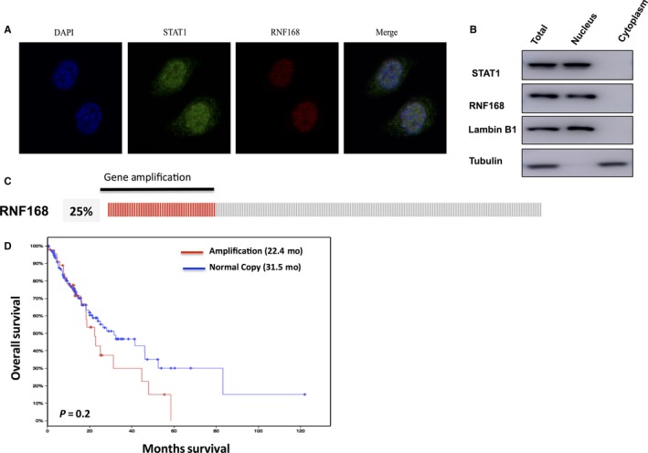
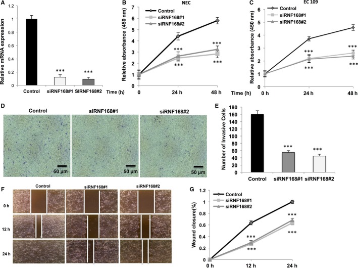
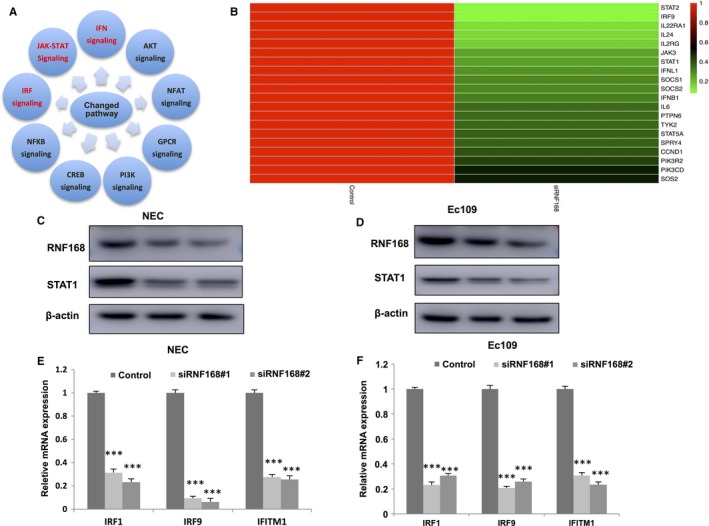
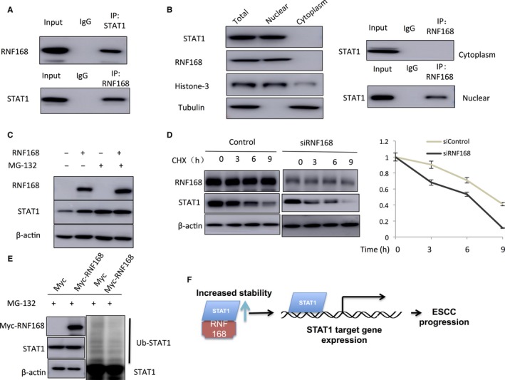
Oesophageal cancer ranks as one of the most common malignancy in China and worldwide. Although genome-wide association studies and molecular biology studies aim to elucidate the driver molecules in oesophageal cancer progression, the detailed mechanisms remain to be identified. Interestingly, RNF168 (RING finger protein 168) shows a high frequency of gene amplification in oesophageal cancer from TCGA database. Here, we report an important function for RNF168 protein in supporting oesophageal cancer growth and invasion by stabilizing STAT1 protein. RNF168 gene is amplified in oesophageal cancer samples, which tends to correlate with poor prognosis. Depletion RNF168 causes decreased cell proliferation and invasion in oesophageal cancer cells. Through unbiased RNA sequencing in RNF168 depleted oesophageal cancer cell, we identifies JAK-STAT pathway is dramatically decreased. Depletion RNF168 reduced JAK-STAT target genes, such as IRF1, IRF9 and IFITM1. Immuno-precipitation reveals that RNF168 associates with STAT1 in the nucleus, stabilizing STAT1 protein and inhibiting its poly-ubiquitination and degradation. Our study provides a novel mechanism that RNF168 promoting JAK-STAT signalling in supporting oesophageal cancer progression. It could be a promising strategy to target RNF168 for oesophageal cancer treatment.

Keywords: RNF168; STAT1; esophageal cancer; stability.


Figures

Figure 1
Figure 1
RNF168 has high frequency of gene amplification in oesophageal cancer and tends to correlate with poor prognosis in oesophageal cancer patients. (A) NEC cells cultured on sterile glass cover slips were fixed with 4% formaldehyde for 10 min. Then the cells were incubated in permeabilization buffer (0.3% Triton X100, in phosphate buffered saline [PBS]) for 10 min. Ten percent donkey serum was added to suppress nonspecific antibody binding at room temperature for 30 min and primary antibodies were incubated overnight at 4°. Fluorochrome‐conjugated secondary antibodies were added after wash in a dark chamber at room temperature. The slides were washed with PBS and mounted using mounting solution containing DAPI. Finally, slides were visualized with a NIKON80i fluorescent microscope. The antibodies were used as follows: anti‐RNF168 (SC‐101125, santa cruz); anti‐STAT1 (9172, Cell Signalling Technology). (B) RNF168 is mainly localized in the nuclear. The subcellular protein fractionation kit (Thermo Scientific, 78840) was used for cytoplasm and nuclear separation. Tubulin and Lambin B1 were used for cytoplasm and nuclear control. (C, D) RNF168 has gene amplification in 25% of esophageal cancer patients. The TCGA database was used to extract the gene expression data. RNF168 gene amplification tends to correlate with poor overall survival in esophageal cancer patients
Figure 2
Figure 2
RNF168 depletion inhibits cell proliferation and invasion in oesophageal cancer cells. (A) RNF168 depletion effect by two different siRNA oligos. NEC cell were transfected with siRNF168 or siControl. After 48 h, RNF168 mRNA levels are determined by real‐time PCR with 36B4 as internal control. (B, C) The WST‐1 assay was used to determine the cellular metabolic activity at indicated time points after transfection. NEC and EC109 cells were transfected with siRNF168 and siControl. After 24 h, cells were seeded into 96‐well plates. These experiments were done in triplicates. All values are mean ± SD (n = 3, *< 0.05; **P < 0.01, ***P < 0.001). (D, E) Transwell invasion assay of NEC cells transfected with indicated RNF168 siRNA. The cell number is counted and data are presented as ±SD. **, P < 0.01, ***, P < 0.001 (Student's t test). (F, G) Wound healing assay of NEC transfected with the indicated siRNA. Quantification of wound closure at the indicated time points. Data are presented as ±SD. **, P < 0.01, ***, P < 0.001 (Student's t test)
Figure 3
Figure 3
RNF168 depletion decreases STAT1 protein level and JAKSTAT target genes in esophageal cancer cells. (A) Top 10 signalling pathways significantly decreased by RNF168 depletion in NEC cells. The pathway‐enrichment analysis was used by the threshold P < 0.001 and fold change >2 to derive regulated genes. RNF168 was depleted by siRNA (mix of siRNF168 #1 and siRNF168 #2) or treated with siControl. After 48 h, the whole mRNA was extracted for RNA sequence analysis. The siControl and siRNF168 were done in triplicates. B: The heat‐map graph shows the JAKSTAT target genes, which is significantly decreased by RNF168 depletion in NEC cells. The significantly regulated genes were overlapped with publish JAKSTAT target gene data. (C, D) RNF168 depletion effect on STAT1 protein level by two different siRNA oligos. NEC and EC109 cells were transfected with siRNF168 or siControl. After 48 h, RNF168 and STAT1 protein levels were determined by Western blot analysis. Actin was used as internal control. (E, F) RNF168 depletion decreases STAT1 target genes using two different siRNA oligos. NEC and EC109 cells were transfected with siRNF168 or siControl. After 48 h, cells, total RNA was prepared and the expression of the endogenous STAT1 target genes, IRF1, IRF9 and IFITM1 were determined by qPCR. Shown are the results from three experiments. *P < 0.05; **P < 0.01; ***P < 0.001 for target gene expression comparison
Figure 4
Figure 4
RNF168 associates with STAT1 in the nuclear and increases STAT1 protein stability. (A) Co‐IP assay reveals association between endogenous RNF168 and STAT1 in NEC cells. NEC cells were harvested with NP‐40 lysis buffer. COIP was performed using antibody as indicated. (B) RNF168 is mainly localized in the nuclear and associates with STAT1 in the nuclear. The subcellular protein fractionation kit (Thermo scientific, 78840) was used for cytoplasm and nuclear separation. Tubulin and Histone‐3 were used for cytoplasm and nuclear control. Based on the separation, IP was done by RNF168 antibody in both the cytosol and nuclear lysis. STAT1 antibody was used to detect the interaction in both the cytosol and nuclear. (C) In the presence of the proteasome inhibitor MG132, the stabilization effect of RNF168 on STAT1 did not further increase STAT1 protein levels. HEK293 cells were transfected with 2 μg STAT1 plasmid and 0.5 μg Myc‐tag or Myc‐RNF168 plasmids. After 24 h, cells were treated with 10 μmol/L MG132/vehicle for 6 h. Cell lysates were prepared for Western blot analysis. The results are representative for three independent experiments. (D) RNF168 increases STAT1 half‐life in NEC cells. NEC cells were transfected with 50 nmol/L siControl or siRNF168 siRNA. After 24 h, cells were treated with 100 μmol/L cycloheximide/vehicle for indicated times. Cell lysates were prepared for Western blot analysis. The results are representative for three independent experiments. The STAT1 relative density was measured by Image J software. (E) RNF168 prohibits STAT1 poly‐ubiquitination. HEK293 cells were transfected with 2 μg STAT1 plasmid and 0.5 μg Myc‐tag or Myc‐RNF168 plasmids. After 24 h, cells were treated with 10 μmol/L MG132 or vehicle for 6 h. Cells were directly harvested and Western blot analysis using STAT1 antibody was used to detect ubiquitinated STAT1 forms. The predicted molecular weight of polyubiquitinated STAT1 is indicated. (F) The hypothetical model for the regulatory mechanism in RNF168 on JAKSTAT1 signalling in esophageal cancer cells: RNF168 associates with STAT1 in the nuclear and subsequently stabilizes STAT1 by inhibiting its polyubiqutination process

References

    1. GBD 2016 Disease and Injury Incidence and Prevalence Collaborators . Global, regional, and national incidence, prevalence, and years lived with disability for 328 diseases and injuries for 195 countries, 1990‐2016: a systematic analysis for the Global Burden of Disease Study 2016. Lancet. 2017;390(10100):1211‐1259. - PMC - PubMed
    1. Chen W, Zheng R, Baade PD, et al. Cancer statistics in China, 2015. CA Cancer J Clin. 2016;66(2):115‐132. - PubMed
    1. Pennathur A, Gibson MK, Jobe BA, Luketich JD. Oesophageal carcinoma. Lancet. 2013;381(9864):400‐412. - PubMed
    1. Gao YB, Chen ZL, Li JG, et al. Genetic landscape of esophageal squamous cell carcinoma. Nat Genet. 2014;46(10):1097‐1102. - PubMed
    1. Thomas SJ, Snowden JA, Zeidler MP, Danson SJ. The role of JAK/STAT signalling in the pathogenesis, prognosis and treatment of solid tumours. Br J Cancer. 2015;113(3):365‐371. - PMC - PubMed

Publication types

MeSH terms

Associated data