. 2019 Mar;67(3):551-565.
doi: 10.1002/glia.23564. Epub 2018 Dec 2.

Augmented astrocyte microdomain Ca2+ dynamics and parenchymal arteriole tone in angiotensin II-infused hypertensive mice

Affiliations

Augmented astrocyte microdomain Ca2+ dynamics and parenchymal arteriole tone in angiotensin II-infused hypertensive mice

Juan Ramiro Diaz et al. Glia. 2019 Mar.

Abstract

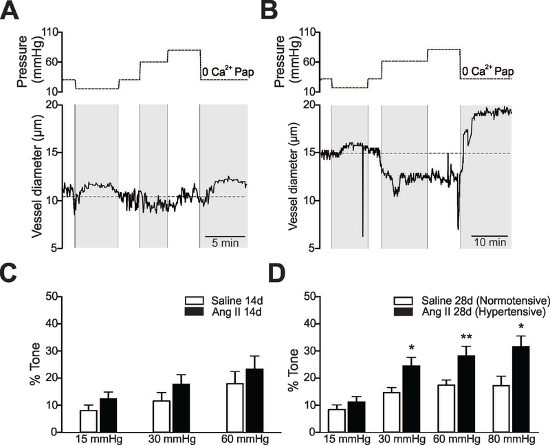
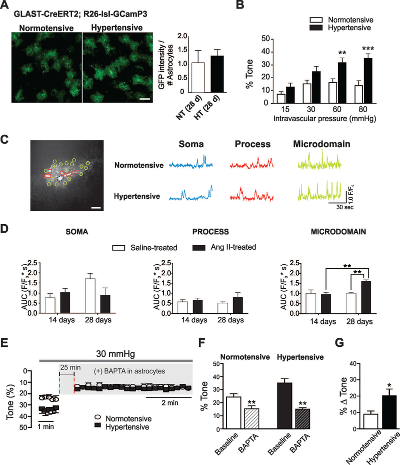
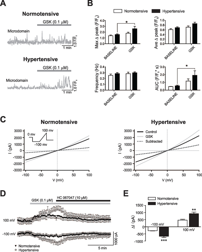
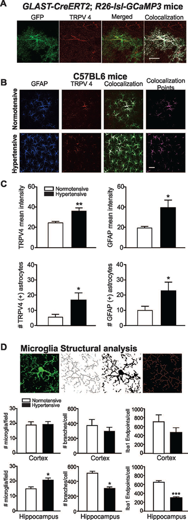
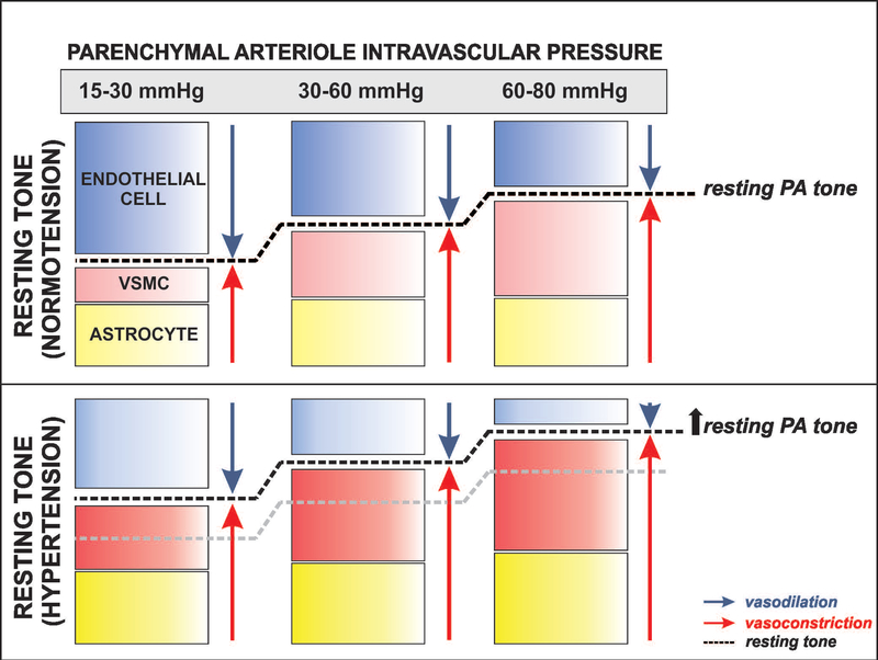
Hypertension is an important contributor to cognitive decline but the underlying mechanisms are unknown. Although much focus has been placed on the effect of hypertension on vascular function, less is understood of its effects on nonvascular cells. Because astrocytes and parenchymal arterioles (PA) form a functional unit (neurovascular unit), we tested the hypothesis that hypertension-induced changes in PA tone concomitantly increases astrocyte Ca2+ . We used cortical brain slices from 8-week-old mice to measure myogenic responses from pressurized and perfused PA. Chronic hypertension was induced in mice by 28-day angiotensin II (Ang II) infusion; PA resting tone and myogenic responses increased significantly. In addition, chronic hypertension significantly increased spontaneous Ca2+ events within astrocyte microdomains (MD). Similarly, a significant increase in astrocyte Ca2+ was observed during PA myogenic responses supporting enhanced vessel-to-astrocyte signaling. The transient potential receptor vanilloid 4 (TRPV4) channel, expressed in astrocyte processes in contact with blood vessels, namely endfeet, respond to hemodynamic stimuli such as increased pressure/flow. Supporting a role for TRPV4 channels in aberrant astrocyte Ca2+ dynamics in hypertension, cortical astrocytes from hypertensive mice showed augmented TRPV4 channel expression, currents and Ca2+ responses to the selective channel agonist GSK1016790A. In addition, pharmacological TRPV4 channel blockade or genetic deletion abrogated enhanced hypertension-induced increases in PA tone. Together, these data suggest chronic hypertension increases PA tone and Ca2+ events within astrocytes MD. We conclude that aberrant Ca2+ events in astrocyte constitute an early event toward the progression of cognitive decline.

Keywords: astrocytes; hypertension; myogenic tone; neurovascular unit; parenchymal arteriole; two to five subject codes.


Figures

Figure 1.
Figure 1.. Hypertension in mice increases parenchymal arteriole tone.
A, B (Upper panels) Pressure protocol applied to induce myogenic responses in cortical parenchymal arterioles (PA). At the end of the experiment, maximum diameter was determined at 30 mmHg upon zero Ca2+/papavarine superfusion. (Bottom panels) Representative PA diameter traces from 28 day saline (A) and Ang II (600 ng/Kg/min) treated (B) C57BL6 mice. (C, D) % PA tone across the pressure range 15–80 mmHg in 14 days (C) (n=8 vessels, 7 saline mice and n=9 vessels, 8 Ang II-treated mice) and 28 days (D) (n=9 vessels, 8 saline mice and n=10 vessels, 9 Ang II treated mice) saline/Ang II-treated mice. Mean ± SEM. Two way ANOVA and Newman-Keuls post-hoc test. *P<0.05, **P<0.01.
Figure 2.
Figure 2.. Hypertension in GLAST-CreERT2; R26-lsl-GCaMP3 mice increases parenchymal arteriole tone and enhances spontaneous Ca2+ oscillations in cortical astrocytes.
(A) Representative images of cortical astrocytes from normotensive and hypertensive GLAST-CreERT2; R26-lsl-GCaMP3 mice labeled with anti-GFP (green) (left). Averaged GFP intensity per astrocyte for both groups (right) (n=12 images from 3 mice), scale bar = 50 μm, unpaired t-test. (B) % parenchymal arteriole (PA) tone across the pressure range 15–80 mmHg, (n=8 vessels, 7 (normotensive (white bar) and 6 hypertensive (black bars) mice). (C) Left, Representative astrocyte image from a GLAST-CreERT2; R26-lsl-GCaMP3 mice showing various regions of interest delineating the cell soma (dashed circle, blue), processes (square, red) and microdomains (MD) (circle, light green), scale bar = 10 μm. Right, Representative traces of spontaneous astrocyte Ca2+ events from soma, processes and MD. (D) Summary data of area under the curve AUC (F/F0*s) for soma, processes and MD from mice treated with saline or Ang II for 14 and 28 days. For saline-treated (white bars) n=10 cells for 14 days and 8 cells for 28 days from 3 and 4 mice, respectively. For Ang II-treated (black bars) n=11 cells for 14 days and 8 cells for 28 days from 3 and 4 mice, respectively. (E) % PA tone from normotensive (open circles) and hypertensive (closed squares) PA before and after introduction of BAPTA into the astrocyte syncytium. (F) Summary of data shown in (E) before (solid bars) and after (dashed bars) Ca2+ chelation. (G) ΔPA arteriole tone from normotensive (white) and hypertensive (black) mice following introduction of BAPTA into astrocytes (n=5 vessels from 4 normotensive and 3 hypertensive mice). Mean ± SEM, Two way ANOVA with Newman-Keuls post-hoc test (for B and D); paired t-test (for F) and unpaired t-test (for G). *P<0.05, **P<0.01 and ***P<0.001.
Figure 3.
Figure 3.. Pressure/flow-evoked astrocyte Ca2+ events in normotensive and hypertensive mice.
(A) Intravascular pressure protocol [15 to 30 mmHg (a), 30 to 60 mmHg (b) and 60 to 80 mmHg (c)] applied to parenchymal arterioles while measuring Ca2+ responses in cortical asrtocytes from GLAST-CreERT2; R26-lsl-GCaMP3 mice. (B-D) Summary data for astrocyte Ca2+ area under the curve (AUC) (F/F0*s) in soma, processes and MD from normotensive (white bars) and hypertensive (black bars) astrocytes in response to intravascular pressure changes (a-c). For protocol a, b: n=6 and 7 cells from 4 mice; for protocol c: n=5 and 5 cells from 3 mice, respectively. Mean ± SEM. Two way ANOVA with Newman-Keuls post-hoc test. *P<0.05.
Figure 4.
Figure 4.. Hypertension enhances TRPV4-mediated Ca2+ activity and whole cell currents in cortical astrocytes.
(A) Representative traces of astrocyte Ca2+ events from microdomains (MD) in response to the TRPV4 channel agonist GSK. (B) Summary data for MD astrocyte Ca2+ parameters (maximum Δpeak (F/F0), average Δpeak (F/F0), frequency (Hz) and area under the curve (AUC) (F/F0*s) from normotensive (white bars, n=9 cells from 3 mice) and hypertensive (black bars, n=11 cells, 3 mice) mice before and after bath applied GSK. (C) Representative whole-cell current traces induced with a voltage ramp protocol (inset) under control (black line) and during GSK bath-application (0.1 μM, light gray line), the subtracted current is shown (dashed line) in normotensive (left) and hypertensive (right) mice. (D) Time course of GSK induced currents obtained at −100 mV and +100mV in normotensive (close triangles) and hypertensive (open circles) mice. (E) Summary data showing Δ currents obtained at −100 mV and +100 mV in normotensive (n=7 cells, 3 mice) (white bars) and hypertensive (n=7 cells, 3 mice) (black bars) mice. Mean ± SEM. Two way ANOVA with Newman-Keuls post-hoc test for figure B and Unpaired T-test for figure D. *P<0.05, ** P<0.01 and *** P<0.0001.
Figure 5.
Figure 5.. Hypertension increases TRPV4 channel expression in astrocytes.
(A) Representative image showing TRPV4 channel (red) expression from a GCaMP3-expressing astrocyte (GFP reporter in green), merged and colocalized images also shown, scale bar = 20 μm. (B) Representative images of brain slices from normotensive and hypertensive mice stained against GFAP (blue) and TRPV4 channel (red), colocalization points (magenta) shown. (C) Summary data for TRPV4 and GFAP mean fluorescence intensity and number of astrocytes in normotensive (white bars) and hypertensive (black bars) mice (n= 9 and 10 images from 6 normotensive and 8 hypertensive mice, respectively). (D) Microglia cell count and structural analysis in cortex and CA1 hippocampus regions (n=3 normotensive and n=3 hypertensive mice). Unpaired T-test, *P<0.05.
Figure 6.
Figure 6.. TRPV4 channel contribution to parenchymal arteriole tone in hypertension.
(A, B) Pressure protocol induced to measure myogenic responses in cortical parenchymal arterioles (PA) in the absence and then presence of the TRPV4 channel blocker HC67047 (10 μM). At the end of the experiment, maximum diameter was determined from values reached in zero Ca2+/papavarine at 30 mmHg. Representative PA diameter traces from normotensive (A) and hypertensive (B) C57BL6 mice (bottom panels). (C, D) Summary data from protocols shown in (A) corresponding to normotensive (C) (n=7 arterioles from 7 animals) and hypertensive (n=7 arterioles from 7 animals) (D) mice subjected to an increase in lumen pressure from 30–60 mmHg in the absence (solid bars) and then presence (hatch bars) of TRPV4 channel blocker. (E) % PA tone before and after introduction of L-NAME (10 μM) and indomethacin (10 μM) to the arteriole lumen followed by bath application (25 min) of HC67047 (10 μM). (F) % PA tone across the pressure range 15–80 mmHg from TRPV4−/− normotensive (white bars) (n=8 vessels, 7 mice) and hypertensive (black bars)(n=8 vessels, 6 mice) mice. Mean ± SEM. For C, D, Paired T-test; for E, One way Anova and Tukey’s multiple comparisons test; for F, Two way ANOVA and Newman-Keuls post-hoc test. *P<0.05, **P<0.01, ****P<0.0001.
Figure 7.
Figure 7.. Proposed model depicting the relative contribution of vascular and non-vascular cells defining resting tone in normotensive and hypertensive parenchymal arterioles.
We propose that at low intravascular pressures (15–30 mmHg) resting parenchymal arteriole (PA) tone is determined by signals from endothelial cells and, to a lesser extent, that of vascular smooth muscle cells (VSMC) and astrocytes. As the intravascular pressure increases to physiological ranges for PA (30–60 mmHg), myogenic constriction and increased astrocyte Ca2+ levels augment resting PA tone. At significantly higher intravascular pressure ranges (60–80 mmHg) myogenic constriction and augmented astrocytes Ca2+ signaling events override endothelial-mediated dilations further increasing PA resting tone. In hypertension, the combined effect of endothelial dysfunction (shown by a lighter blue color) with augmented VSMC constriction and resting astrocyte Ca2+ (shown with darker red and yellow colors, respectively) shifts the relative contribution of these three players (endothelium, VSMC and astrocytes) favoring constriction and resulting in higher levels of resting PA tone.

References

    1. Agarwal A, Wu PH, Hughes EG, Fukaya M, Tischfield MA, Langseth AJ, Wirtz D, Bergles DE. 2017. Transient Opening of the Mitochondrial Permeability Transition Pore Induces Microdomain Calcium Transients in Astrocyte Processes. Neuron 93:587–605 e7. - PMC - PubMed
    1. Attwell D, Buchan AM, Charpak S, Lauritzen M, Macvicar BA, Newman EA. 2010. Glial and neuronal control of brain blood flow. Nature 468:232–43. - PMC - PubMed
    1. Bagher P, Beleznai T, Kansui Y, Mitchell R, Garland CJ, Dora KA. 2012. Low intravascular pressure activates endothelial cell TRPV4 channels, local Ca2+ events, and IKCa channels, reducing arteriolar tone. Proc Natl Acad Sci U S A 109:18174–9. - PMC - PubMed
    1. Baumbach GL, Heistad DD. 1989. Remodeling of cerebral arterioles in chronic hypertension. Hypertension 13:968–72. - PubMed
    1. Baylie RL, Brayden JE. 2011. TRPV channels and vascular function. Acta Physiol (Oxf) 203:99–116. - PMC - PubMed

Publication types