

Spinal cord and infratentorial lesions in radiologically isolated syndrome are associated with decreased retinal ganglion cell/inner plexiform layer thickness
- PMID: 30507269
- PMCID: PMC6546560
- DOI: 10.1177/1352458518815597
Spinal cord and infratentorial lesions in radiologically isolated syndrome are associated with decreased retinal ganglion cell/inner plexiform layer thickness
Abstract
Background: The role of retinal imaging with optical coherence tomography (OCT) in assessing individuals with radiologically isolated syndrome (RIS) remains largely unexplored.
Objective: To assess retinal layer thicknesses in RIS and examine their associations with clinical features suggestive of increased risk for conversion to multiple sclerosis (MS).
Methods: A total of 30 RIS subjects and 60 age- and sex-matched healthy controls (HC) underwent retinal imaging with spectral-domain OCT, followed by automated segmentation of retinal layers.
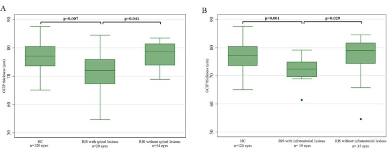
Results: Overall, retinal layer thicknesses did not differ between RIS and HC. However, RIS subjects with spinal cord (SC) lesions had lower ganglion cell + inner plexiform layer (GCIP) thickness compared to HC (-4.41 μm; p = 0.007) and RIS without SC lesions (-3.53 μm; p = 0.041). Similarly, RIS subjects with infratentorial (IT) brain lesions had lower GCIP thickness compared to HC (-4.07 μm; p < 0.001) and RIS without IT lesions (-3.49 μm; p = 0.029). Multivariate analyses revealed that the presence of SC or IT lesions were independently associated with lower GCIP thickness in RIS (p = 0.04 and p = 0.03, respectively). Other patient characteristics, including sex, abnormal cerebrospinal fluid, and presence of gadolinium-enhancing or juxtacortical lesions, were not associated with retinal layer thicknesses.
Conclusion: The presence of SC or IT lesions in RIS may be associated with retinal neuro-axonal loss, supporting the presence of more disseminated disease.
Keywords: Multiple sclerosis; demyelination; optical coherence tomography; radiologically isolated syndrome; retina; spinal cord.
Conflict of interest statement
Conflict of interest:
The authors declare that there is no conflict of interest.
Figures
References
-
- Okuda DT, Mowry EM, Beheshtian A, et al. Incidental MRI anomalies suggestive of multiple sclerosis: the radiologically isolated syndrome. Neurology 2009; 72: 800–805. - PubMed
-
- Kantarci OH, Lebrun C, Siva A, et al. Primary Progressive Multiple Sclerosis Evolving From Radiologically Isolated Syndrome. Annals of Neurology 2016; 79: 288–294. - PubMed
-
- Lebrun C, Bensa C, Debouverie M, et al. Association Between Clinical Conversion to Multiple Sclerosis in Radiologically Isolated Syndrome and Magnetic Resonance Imaging, Cerebrospinal Fluid, and Visual Evoked Potential: Follow-up of 70 Patients. Archives of Neurology 2009; 66: 841–846. - PubMed
Publication types
MeSH terms
Grants and funding
LinkOut - more resources
Full Text Sources
