Review
. 2018;66(4):1341-1362.
doi: 10.3233/JAD-180549.

The Role of Cognitive Reserve in Alzheimer's Disease and Aging: A Multi-Modal Imaging Review

Affiliations
Review

The Role of Cognitive Reserve in Alzheimer's Disease and Aging: A Multi-Modal Imaging Review

Arianna Menardi et al. J Alzheimers Dis. 2018.

Abstract

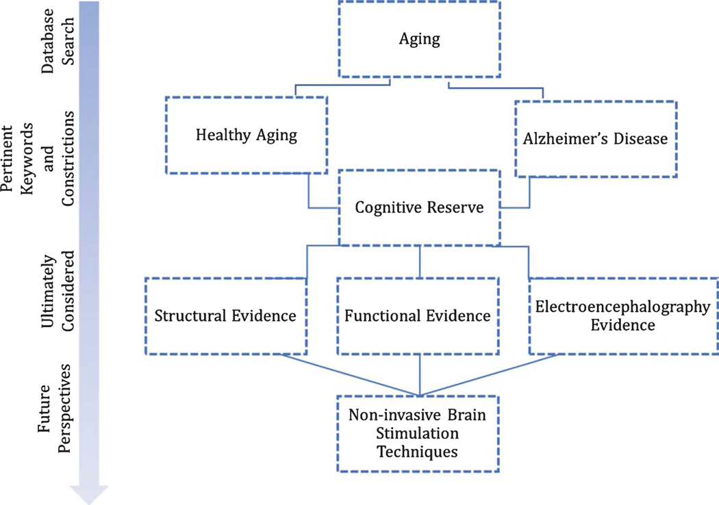
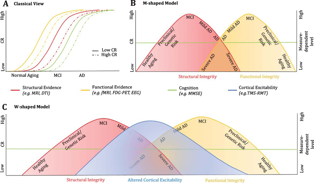
Comforts in modern society have generally been associated with longer survival rates, enabling individuals to reach advanced age as never before in history. With the increase in longevity, however, the incidence of neurodegenerative diseases, especially Alzheimer's disease, has also doubled. Nevertheless, most of the observed variance, in terms of time of clinical diagnosis and progression, often remains striking. Only recently, differences in the social, educational and occupational background of the individual, as proxies of cognitive reserve (CR), have been hypothesized to play a role in accounting for such discrepancies. CR is a well-established concept in literature; lots of studies have been conducted in trying to better understand its underlying neural substrates and associated biomarkers, resulting in an incredible amount of data being produced. Here, we aimed to summarize recent relevant published work addressing the issue, gathering evidence for the existence of a common path across research efforts that might ease future investigations by providing a general perspective on the actual state of the arts. An innovative model is hereby proposed, addressing the role of CR across structural and functional evidences, as well as the potential implementation of non-invasive brain stimulation techniques in the causal validation of such theoretical frame.

Keywords: Aging; Alzheimer’s disease; cognitive reserve; diffusion tensor imaging; electroencephalography; functional magnetic resonance imaging; magnetic resonance imaging; positron emission tomography; transcranial magnetic stimulation.


Figures

Fig. 1.
Fig. 1.
Schematic flowchart of the literature search and rationale behind the study.
Fig. 2.
Fig. 2.
Associations between structural measures and CR in healthy and pathological brain aging. A) In healthy aging, a positive association has been reported between grey matter volume and CR, both at the whole brain level, as well as within specific regions, i.e., mainly the fronto-parietal sites [51]. In contrast, a negative correlation between CR and measures of FA in the genu of the corpus callosum—a region showing great physiological changes with age—has been reported [51]. According to the authors, the observed negative, rather than positive, relationship may reflect “a greater capacity to tolerate age-related structural damage without concomitant impact on cognitive performance,” In other words, individuals at preclinical/genetic risk conditions, who nonetheless have high CR, could lose white matter integrity without manifesting cognitive impairment, thus representing a transitional or intermediate brain status between healthy and pathological aging. Overall, grey and white matter changes support the continuous protective role of CR in sustaining preserved functioning for longer during time. B) In contrast with healthy cognitive aging, MCI and AD show negative associations between CR and grey matter volume at the global and regional level. In particular, the hippocampal formation represents one of the regions showing the greatest atrophy as a function of reserve, which suggests CR may enable patients to sustain more advanced stages of pathology compared to their low CR counterpart. A similar pattern has been observed in terms of FA reduction over pathologically relevant fibers’ tracts, including the corpus callosum, the cingulum, the inferior and superior longitudinal fasciculi and the fronto-occipital bundle [51], with a greater negative slope in MCI compared to AD patients [51].
Fig. 3.
Fig. 3.
Task-associated functional activation/deactivation and CR in healthy and pathological cognitive aging. Task-induced activation and deactivation in regions showing greatest difference in the regression slope between healthy subjects and the pathological sample [89]. As a function of CR, healthy elderlies show a decrease in the recruitment of regions active during the task at hand (in this case, language related areas- middle/superior frontal gyrus and anterior cingulate cortex mainly), and reduced deactivation in some of the regions belonging to the DMN (precuneus, posterior cingulate cortex). Such finding suggests higher CR is associated with a more efficient allocation of metabolic resources and information processing. On the other hand, AD patients need greater activation to carry out the same task, thus proving better compensatory capacities as a function of CR. Greater reduction is observed in regions typically showing task-induced deactivations, i.e., DMN regions. Amnestic-MCI patients show less dramatic effects; a tendency mirroring that of the AD sample can still be appreciated.
Fig. 4.
Fig. 4.
Proposed model of cortical excitability as a function of disease progression and cognitive performance. Lower RMT (i.e., higher cortical excitability) has been observed in pathological cognitive aging compared to healthy cohorts, reflecting a possible compensatory mechanism for the progressive loss of cortical neurons. We speculate that the decrease in the RMT would be observed until the most advanced stages of the disease when a critical point in cortical thinning is reached. In line with this notion, higher cortical excitability is associated with better cognitive performance in young subjects and to progressively shift toward a negative association at the earliest stages of AD. The progressive change in slope between cortical excitability and cognitive performance is thought to reflect the point of inflection where hyperexcitability no longer acts as a compensatory mechanism, but rather starts to become detrimental for the subject, possibly reflecting a greater impediment in cognitive resource allocation. CR is hereby proposed to ensure preserved functioning for longer during time, delaying the time when the inflection point is reached by the individual.
Fig. 5.
Fig. 5.
Contrasting models of the impact of CR on the manifestation and progression of pathological brain states. The traditional view on the role of CR (A) hypothesizes that CR simply acts in protecting the individual, delaying the time of symptomatology onset and of clinical diagnosis. On the other hand, a M-shaped model (B) reflexes a much more complex reality in which not only healthy subjects and pathological individuals show gradual opposite changes in slope as a function of CR, but also structural and functional modifications show mirror-like behavior. In the proposed model, the term structural integrity refers to the collected evidence of global and regional changes in the cerebral volume, such as that, in healthy aging, higher reserve correlates with more robust neural substrates, i.e., greater grey matter volumes and higher measures of FA in the connecting fibers. On the other hand, a progressive shift in slope is observed in the pathological sample (from MCI to AD), where higher CR becomes associated with thinner cortices, providing its compensatory role in sustaining greater degree of severity for longer in time. Similarly, functional integrity refers to the reported changes in the functional recruitment of regions that ensure the individual to maintain an appropriate level of cognitive functioning. In respect to CR, healthy elderlies show less functional activation over task-relevant regions and reduced deactivation of the DMN (not shown in the model; see Fig. 3), proving more finely tuned patterns of activity and effortless processing. On the other hand, in the AD pathology, CR acts in ensuring greater functional activation and recruitment of adjacent regions (mainly frontal areas), to maintain an efficient cognitive profile despite disease progression. The reported slope change in functional activity represent therefore task-driven, more than resting state, accommodations as a function of CR, with similar patterns reported in both fMRI and FDG-PET studies [156]. Notably, preclinical stages of disease or genetic conditions present intermediate brain statuses as a function of CR between healthy and pathological aging. C) represents a W-shaped model, where the altered cortical excitability is seen as the most easily and non-invasive assessable measure summarizing both structural and functional evidences, further proving the potential implementation of NIBS in clinical practice in the evaluation of the individual collocation along the healthy-pathological axis.

References

    1. Alzheimer’s Association (2017) 2017 Alzheimer’s disease facts and figures. Alzheimers Dement 13, 325–373.
    1. Driscoll I, Resnick SM, Troncoso JC, An Y, O’Brien R, Zonderman AB (2006) Impact of Alzheimer’s pathology on cognitive trajectories in nondemented elderly.AnnNeurol 60, 688–695. - PubMed
    1. Snowdon DA (2003) Healthy aging and dementia: Findings from the Nun Study. Ann Intern Med 139, 450–454. - PubMed
    1. Satz P (1993) Brain reserve capacity on symptom onset after brain injury: A formulation and review of evidence for threshold theory. Neuropsychology 7, 273–295.
    1. Katzman R (1993) Education and the prevalence of dementia and Alzheimer’s disease. Neurology 43, 13–20. - PubMed

Publication types