. 2019 Jan 2;129(1):310-323.
doi: 10.1172/JCI95731. Epub 2018 Dec 3.

Chemogenetic silencing of hippocampal neurons suppresses epileptic neural circuits

Affiliations

Chemogenetic silencing of hippocampal neurons suppresses epileptic neural circuits

Qi-Gang Zhou et al. J Clin Invest. .

Abstract

We investigated how pathological changes in newborn hippocampal dentate granule cells (DGCs) lead to epilepsy. Using a rabies virus-mediated retrograde tracing system and a designer receptors exclusively activated by designer drugs (DREADD) chemogenetic method, we demonstrated that newborn hippocampal DGCs are required for the formation of epileptic neural circuits and the induction of spontaneous recurrent seizures (SRS). A rabies virus-mediated mapping study revealed that aberrant circuit integration of hippocampal newborn DGCs formed excessive de novo excitatory connections as well as recurrent excitatory loops, allowing the hippocampus to produce, amplify, and propagate excessive recurrent excitatory signals. In epileptic mice, DREADD-mediated-specific suppression of hippocampal newborn DGCs dramatically reduced epileptic spikes and SRS in an inducible and reversible manner. Conversely, specific activation of hippocampal newborn DGCs increased both epileptic spikes and SRS. Our study reveals an essential role for hippocampal newborn DGCs in the formation and function of epileptic neural circuits, providing critical insights into DGCs as a potential therapeutic target for treating epilepsy.

Keywords: Epilepsy; Neuronal stem cells; Neuroscience; Stem cells.


Conflict of interest statement

Conflict of interest: The authors have declared that no conflict of interest exists.

Figures

Figure 1
Figure 1. Inhibition of hippocampal DGCs suppresses SRS.
(A) hM4Di and YFP are expressed in hippocampal DGCs in POMC-Cre;hM4Difl/+;YFPfl/+ mice. (B) CRE is expressed in the DG, and YFP, which is an indicator of hM4Di, is expressed in all DGCs in POMC-Cre;hM4Difl/+;YFPfl/+ mice. (C) Representative EEG traces in the absence and presence of CNO in POMC-Cre;hM4Difl/+ mice during pilocarpine-induced epilepsy. (D and E) On days 1–3, vehicle treatment did not significantly change epileptic spikes or SRS in either control or POMC-Cre;hM4Difl/+ mice. On days 4–6, CNO-mediated suppression of hippocampal DGCs significantly reduced epileptic spikes (SPKs ) (n = 19) as well as SRS (n = 11) in POMC-Cre;hM4Difl/+ mice in an inducible and reversible manner, as determined by 2-way RM ANOVA with a Bonferroni’s multiple comparison post test. On days 7–9, epileptic spikes and SRS returned to basal levels in POMC-Cre;hM4Difl/+, showing CNO-dependent transient and reversible suppression of DGC activity. Note that epilepsy spikes and SRS were quantified during the 24 hours after vehicle (days 1–3, blue circles), CNO (days 4-6, red circles), and recovery without treatment (days 7-9, green circles). Two-way RM ANOVA with Bonferroni’s multiple comparison tests were used for D and E. Asterisks indicate that CNO treatment resulted in a significant reduction in epileptic spikes and SRS compared with vehicle treatment, which returned to basal levels during the recovery period. ***P < 0.001.
Figure 2
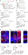
Figure 2. Aberrant integration of DGCs into proepileptic neural circuits depends upon the age of DGCs.
(A) Experimental schematic showing mapping of neuronal connectivity of DGCs born before and after SE induction. (B) The connectivity ratio of input neurons to starter cells shows the critical time window during which newborn DGCs of different ages are recruited to proepileptic neural circuits (n = 4, each). (CF) Connectivity ratios of DGCs born 3 and 14 days after SE and 7 and 21 days before SE are shown. DGCs born between 7 days before and 3 days after SE induction show aberrant connectivity, whereas the connectivity of DGCs born 21 days before or 14 days after SE induction is not significantly different (n = 4, each). (GJ) DGCs born 3 days after SE, but not those born 21 days prior to SE, are ectopically located, are hypertrophic, and develop hilar basal dendrites (n = 4, each). Arrows (G and H) indicate disorganized projections. Disorganized axonal projections were evident when the input of DGCs born 3 days after, but not 21 days before, SE induction was examined. Red lines on the right side of each image (I) indicate the location of projections. *P < 0.05; ***P < 0.001, 2-way ANOVA with Bonferroni’s multiple comparison tests (B) and Mann-Whitney nonparametric U tests (CF).
Figure 3
Figure 3. Aberrant integration of hippocampal DGCs contributes to the formation of recurrent excitatory loops.
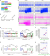
(A) Experimental schematic showing the experimental design for rabies virus–mediated retrograde tracing in epileptic mice. (BD) The connectivity ratio to input neurons of DGCs born 3 days after SE was significantly increased (n = 4 each). (E) Graphs show increases in connectivity ratios between input neurons located in subregions of major brain structures and newborn DGCs (n = 4 each). (F) Representative images showing significant changes in the number of input neurons that directly connected to newborn DGCs in epileptic mice. (G) The connectivity ratio was proportional to the severity of epilepsy, as assessed by the frequency of epileptic spikes and SRS (n = 10). Data represent mean ± SEM. *P < 0.05, Mann-Whitney U tests (BE) and Pearson’s correlation (G).
Figure 4
Figure 4. The essential role of hippocampal DGCs in the production of seizures during epilepsy.
(A and B) Experimental schematics showing the strategy to selectively suppress the neuronal activity of hippocampal newborn DGCs in NCE;hM4Difl/+ or NCE;hM4Difl/+;YFPfl/+ mice. (C) Representative EEG recordings show that CNO treatment suppressed epileptic spikes as well as SRS in NCE;hM4Difl/+mice, but not in hM4Difl/+ control mice. (D and E) Epileptic spikes and SRS were quantified during 24 hours of vehicle (days 1–3, blue circles), CNO (days 4–6 and 10, red circles), and recovery without treatment (days 7–9 and 11, green circles). Quantitative results show that CNO treatment effectively reduced the frequency of epileptic spikes (n = 17) and SRS (n = 9) only in NCE;hM4Difl/+ mice during epilepsy. ***P < 0.001. (F and G) Percentage of inhibition of epileptic spikes was proportional to the total number of hM4Di-expressing YFP-positive cells, as well as aberrant hM4Di-expressing YFP-positive cells, in the dentate gyrus. Asterisks in D and E indicate that CNO treatment resulted in a significant reduction in epileptic spikes and SRS compared with vehicle treatment, which returned to basal levels during each recovery period. Two-way RM ANOVA with Bonferroni’s multiple comparison tests (D, E) or Pearson’s correlation (F, G) were used.
Figure 5
Figure 5. The requirement of adult-born DGCs for the expression of seizures in epileptic mice.
(A) Experimental schematic showing strategy for hM4Di receptor expression exclusively in newborn DGCs using RV-hM4Di-IRES-GFP. (B) The process of vehicle treatment (blue circles), CNO treatment (red circles), and recovery without treatment (green circles) was performed on days 1, 2, and 3, respectively. Epileptic spikes and SRS were quantified for 8 hours per day on days 1–3. CNO treatment effectively reduced the occurrence of epileptic spikes and SRS in mice injected with RV-hM4Di-IRES-GFP, but not with RV-GFP. Twenty-four hours after CNO treatment, epileptic spikes and SRS returned to basal levels in RV-hM4Di-IRES-GFP mice. (C) Level of inhibition of epileptic spikes was proportional to the number of hM4Di-expressing YFP-positive cells. **P < 0.01; ***P < 0.001, 2-way RM ANOVA with Bonferroni’s multiple comparison tests (B) and Pearson’s correlation (C).
Figure 6
Figure 6. Specific activation of newborn DGCs is sufficient to induce seizures in epileptic mice.
(A) Schematic showing the experimental plan to activate hippocampal newborn DGCs in epileptic mice. Representative EEG traces show that specific activation of hippocampal newborn DGCs significantly increased EEG activity and epilepsy frequency. (B) Selective activation of hippocampal newborn DGCs dramatically increased epileptic spikes as well as SRS in epileptic mice. (C) CNO-mediated control of neuronal activity was transiently inducible and reversible. Twenty-four hours after the previous CNO-mediated activation of DGCs, repeated CNO administration increased epileptic spikes and SRS. *P < 0.05; ***P < 0.001, 2-way RM ANOVA with Bonferroni’s multiple comparison tests (B) and Wilcoxon’s matched-pairs signed rank tests (C).
Figure 7
Figure 7. Seizure expression increases proportionally to the number of aberrantly developed hippocampal newborn DGCs.
(A and B) Newborn DGCs activated by the CNO-hM3Dq interaction show near complete coexpression with c-FOS in both control (non SE) and epileptic (SE) mice. (C) GFP-labeled hippocampal newborn DGCs develop abnormally in epileptic mice. (D) The number of abnormally developed hippocampal DGCs positively correlates with the severity of epileptic phenotypes, such as epileptic spikes and SRS. Data are shown as mean ± SEM. *P < 0.05; **P < 0.01, Mann-Whitney U tests (A and B) and Pearson’s correlation (D).
Figure 8
Figure 8. De novo formation of excessive excitatory networks and recurrent excitatory loops in the hippocampus of epileptic mice.
In the normal hippocampus (top panel), input neurons located in the EC send their excitatory projections to the dentate gyrus via the perforant pathway. DGCs in the dentate gyrus project their axons (mossy fibers) to CA3 pyramidal neurons. Pyramidal neurons are interconnected, forming recurrent networks in CA3, and project to CA1 neurons via Schaffer collaterals. This unidirectional excitatory neuronal network, cortex→dentate gyrus→CA3→CA1, has been referred to as trisynaptic connections. In epileptic mice (lower panel), excitatory inputs from the EC to the dentate gyrus are increased, reinforcing the convergence of excitatory signals to the dentate gyrus (no. 1). In addition, de novo formation of recurrent excitatory loops within the dentate gyrus (no. 2), as well as between the dentate gyrus and CA3 (no. 3), facilitates the production, amplification, and propagation of excitatory signals and may be responsible for synchronous discharges in the hippocampus.

References

    1. French JA, et al. Characteristics of medial temporal lobe epilepsy: I. Results of history and physical examination. Ann Neurol. 1993;34(6):774–780. doi: 10.1002/ana.410340604. - DOI - PubMed
    1. Williamson PD, et al. Characteristics of medial temporal lobe epilepsy: II. Interictal and ictal scalp electroencephalography, neuropsychological testing, neuroimaging, surgical results, and pathology. Ann Neurol. 1993;34(6):781–787. doi: 10.1002/ana.410340605. - DOI - PubMed
    1. Engel J. Etiology as a risk factor for medically refractory epilepsy: a case for early surgical intervention. Neurology. 1998;51(5):1243–1244. doi: 10.1212/WNL.51.5.1243. - DOI - PubMed
    1. Kwan P, Brodie MJ. Early identification of refractory epilepsy. N Engl J Med. 2000;342(5):314–319. doi: 10.1056/NEJM200002033420503. - DOI - PubMed
    1. Chang BS, Lowenstein DH. Epilepsy. N Engl J Med. 2003;349(13):1257–1266. doi: 10.1056/NEJMra022308. - DOI - PubMed

Publication types