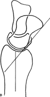
In Brief: The Lichtman Classification for Kienböck Disease
- PMID: 30507834
- PMCID: PMC6554122
- DOI: 10.1097/CORR.0000000000000595
In Brief: The Lichtman Classification for Kienböck Disease
Conflict of interest statement
All ICMJE Conflict of Interest Forms for authors and
Figures


Similar articles
-
The Role of Wrist Arthroscopy in Kienbock Disease.Hand Clin. 2017 Nov;33(4):727-734. doi: 10.1016/j.hcl.2017.07.003. Hand Clin. 2017. PMID: 28991584 Review.
-
The Lichtman classification for Kienböck's disease: an assessment of reliability.J Hand Surg Am. 2003 Jan;28(1):74-80. doi: 10.1053/jhsu.2003.50035. J Hand Surg Am. 2003. PMID: 12563641
-
Is There a Correlation between the Radiological and Clinical Outcome after Core Decompression of the Radius for Kienböck Disease?J Hand Surg Asian Pac Vol. 2024 Feb;29(1):36-42. doi: 10.1142/S2424835524500061. J Hand Surg Asian Pac Vol. 2024. PMID: 38299239
-
Robert Kienbock: the man and his work.J Hand Surg Eur Vol. 2010 Sep;35(7):534-7. doi: 10.1177/1753193410367708. Epub 2010 Apr 28. J Hand Surg Eur Vol. 2010. PMID: 20427409
-
Kienböck Disease: Moving Forward.J Hand Surg Am. 2016 May;41(5):630-8. doi: 10.1016/j.jhsa.2016.02.013. Epub 2016 Apr 5. J Hand Surg Am. 2016. PMID: 27055625 Review.
Cited by
-
Detecting Avascular Necrosis of the Lunate from Radiographs Using a Deep-Learning Model.J Imaging Inform Med. 2024 Apr;37(2):706-714. doi: 10.1007/s10278-023-00964-0. Epub 2024 Jan 16. J Imaging Inform Med. 2024. PMID: 38343256 Free PMC article.
-
Recent Advances in Assessment and Treatment in Kienböck's Disease.J Clin Med. 2022 Jan 27;11(3):664. doi: 10.3390/jcm11030664. J Clin Med. 2022. PMID: 35160115 Free PMC article. Review.
-
Comparing the Radiologic and Functional Outcome of Radial Shortening Versus Capitate Shortening in Management of Kienböck's Disease.Hand (N Y). 2023 Oct;18(7):1120-1128. doi: 10.1177/15589447221081564. Epub 2022 Mar 23. Hand (N Y). 2023. PMID: 35321588 Free PMC article. Clinical Trial.
-
A case report of effective intra-articular elcatonin administration in a patient with osteonecrosis of the lunate.Int J Surg Case Rep. 2023 Apr;105:108056. doi: 10.1016/j.ijscr.2023.108056. Epub 2023 Mar 27. Int J Surg Case Rep. 2023. PMID: 37001370 Free PMC article.
-
Application of 3 dimension-printed injection-molded polyether ether ketone lunate prosthesis in the treatment of stage III Kienböck's disease: A case report.World J Clin Cases. 2022 Aug 26;10(24):8761-8767. doi: 10.12998/wjcc.v10.i24.8761. World J Clin Cases. 2022. PMID: 36157814 Free PMC article.
References
-
- Alexander AH, Turner MA, Alexander CE, Lichtman DM. Lunate silicone replacement arthroplasty for Kienböck’s disease: a long-term follow-up. J Hand Surg Am. 1990;5:401-407. - PubMed
-
- Allan CA, Joshi A, Lichtman DM. Kienböck’s disease: diagnosis and treatment. J Am Acad Orthop Surg . 2001;9:128-136. - PubMed
-
- Bain GI, Begg M. Arthroscopic assessment and classification of Kienböck’s disease. Tech Hand Up Extrem Surg. 2006;10:8-13. - PubMed
-
- Divelbiss B, Baratz ME. Kienböck disease. J Hand Surg Am. 2001;1:61-72.
-
- Goldfarb CA, Hsu J, Gelberman RH, Boyer MI. The Lichtman classification for Kienböck’s disease: an assessment of reliability. J Hand Surg Am. 2003;28:74-80. - PubMed
Publication types
MeSH terms
Personal name as subject
- Actions
LinkOut - more resources
Full Text Sources
Medical

