

Isolation and characterization of mesenchymal stem/stromal cells from Ctenomys minutus
- PMID: 30508008
- PMCID: PMC6415614
- DOI: 10.1590/1678-4685-GMB-2018-0012
Isolation and characterization of mesenchymal stem/stromal cells from Ctenomys minutus
Abstract
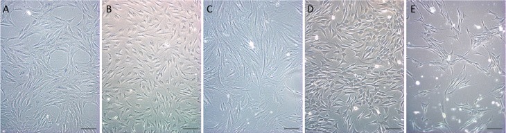
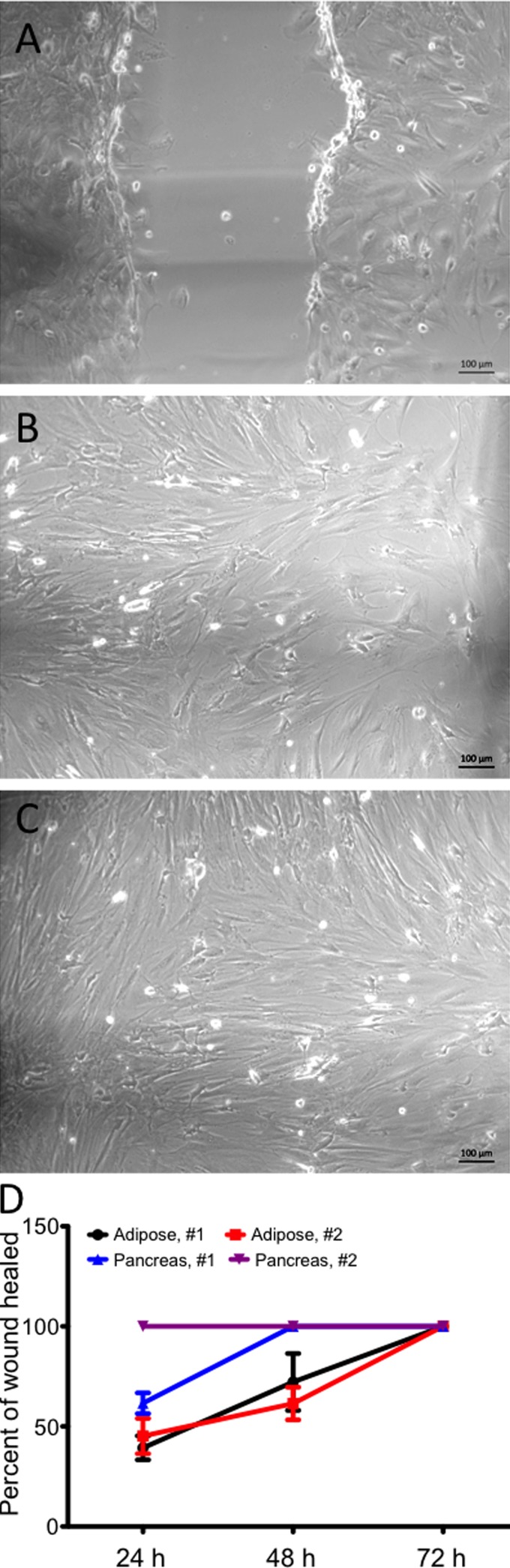
Mesenchymal stem/stromal cells (MSCs) are multipotent cells distributed in all tissues and characterized by adherence, morphology, immunophenotype and trilineage differentiation potential. The present study aimed to isolate and characterize adherent MSC-like populations from different tissues of Ctenomys minutus, a threatened wildlife rodent popularly known as tuco-tuco. Adherent cells were isolated from bone marrow, brain, liver, pancreas and adipose tissue of three adult animals collect in southern Brazil. Cultures showed typical morphology and proliferation potential. Adipose-derived MSCs showed trilineage potential. Cultures derived from adipose tissue, bone marrow and brain were immunophenotyped with negative results for CD31, CD44, CD45, CD106, and MHC class II, as well as strong positive results for CD29. Low fluorescence levels were seen for CD49d, CD90.2 and CD117. Cultures were negative for CD49e, except for brain-derived cultures that were weakly positive. CD11b was negative in adipose-derived MSCs, but positive in brain and bone marrow-derived cultures. The scratch assay showed high migration potential for pancreas and adipose tissue-derived cells. This study represents the first report of isolation and characterization of cultures having characteristics of MSCs from Ctenomys minutus. The collection of biological information for biobanks represents an important contribution to the creation of strategies for prevention of loss of genetic diversity.
Figures


References
-
- Argôlo NM, Neto, Feitosa MLT, Silva SS, Fernandes PB, Pessoa GT, Bezerra DO, Almeida HM, Carvalho YKP, Rocha AR, Silva LMC, Carvalho MAM. Isolation, expansion, differentiation and growth kinetics essay in mesenchymal stem cells culture from the bone marrow of collared peccaries (Tayassu tajacu) Acta Scient Veter. 2016;44:1–11.
-
- Baksh D, Davies JE, Zandstra PW. Adult human bone marrow-derived mesenchymal progenitor cells are capable of adhesion-independent survival and expansion. Exp Hematol. 2003;31:723–732. - PubMed
-
- Bidau CJ. Family Ctenomyidae Lesson, 1842. In: Patton JL, Pardinas UFJ, D’Elia G, editors. Mammals of South America. The University of Chicago Press; Chicago: 2015. pp. 818–877.
Internet Resources
-
- Roth V. Doubling time computing. 2006. http://www.doubling-time/compute.php.
LinkOut - more resources
Full Text Sources
Research Materials
Miscellaneous
