. 2019 Apr 1;76(4):462-469.
doi: 10.1001/jamaneurol.2018.4316.

Association of Seizure Spread With Surgical Failure in Epilepsy

Affiliations

Association of Seizure Spread With Surgical Failure in Epilepsy

John P Andrews et al. JAMA Neurol. .

Abstract

Importance: Seizures recur in as many as half of patients who undergo surgery for drug-resistant temporal lobe epilepsy (TLE). Understanding why TLE is resistant to surgery in some patients may reveal insights into epileptogenic networks and direct new therapies to improve outcomes.

Objective: To characterize features of surgically refractory TLE.

Design, setting, and participants: Medical records from a comprehensive epilepsy center were retrospectively reviewed for 131 patients who received a standard anteromedial temporal resection by a single surgeon from January 1, 2000, to December 31, 2015. Thirteen patients were excluded for having less than 1 year of follow-up. Patients at the highest risk for seizure recurrence were identified. Intracranial electroencephalogram (iEEG) analyses generated 3-dimensional seizure spread representations and quantified rapid seizure spread. The final analyses of seizure outcome and follow-up data were performed in June 2017.

Main outcomes and measures: The Engel class seizure outcome following surgery was evaluated for all patients, defining seizure recurrence as Engel class II or greater. Intracranial recordings of neocortical grids/strips and depth electrodes were analyzed visually for seizure spread. Fast β power was projected onto reconstructions of patients' brain magnetic resonance imaging scans to visualize spread patterns and was quantified to compare power within vs outside resective margins.

Results: Of 118 patients with 1 year of follow-up or more (mean [SD], 6.5 [4.6] years), 66 (55.9%) were women and 52 (44.1%) were men (median age, 39 years [range, 4-66 years]). The cumulative probability of continuous Engel class I seizure freedom since surgery at postoperative year 10 and afterward was 65.6%, with 92% of recurrences in years 1 to 3. Multivariable statistical analyses found that the selection for iEEG study was the most reliable predictor of seizure recurrence, with a mixed-effects model estimating that the Engel score in the iEEG cohort was higher by a mean (SD) of 1.1 (0.33) (P = .001). In patients with iEEG results, rapid seizure spread in less than 10 seconds was associated with recurrence (hazard ratio, 5.99; 95% CI, 1.7-21.1; P < .01). In the first 10 seconds of seizures, fast β power activity outside the resective margins in the lateral temporal cortex was significantly greater in patients whose seizures recurred compared with patients who were seizure-free (mean [SEM], 137.5% [16.8%] vs 93.4% [4.6%]; P < .05).

Conclusions and significance: Rapid seizure spread outside anteromedial temporal resection resective margins plays a significant role in the surgical failure of drug-resistant TLE. Seizure control after epilepsy surgery might be improved by investigating areas of early spread as candidates for resection or neuromodulation.


Conflict of interest statement

Conflict of Interest Disclosures: None reported.

Figures

Figure 1.
Figure 1.. Engel Class I Seizure Freedom
Seizure outcome after isolated anteromedial temporal resection. The cumulative probability of continuous seizure freedom is 83.9%, 77.8%, 70.9%, and 65.6% at 1, 2, 3, and 10 years or more after surgery, respectively.
Figure 2.
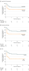
Figure 2.. Predictors of Seizure Recurrence
Analyses of Engel class I seizure freedom since surgery. A-C, Patients with a discordance of preoperative positron emission tomography (PET) with preoperative scalp electroencephalogram (EEG) (A) were significantly more likely to have seizure recurrence (hazard ratio [HR], 2.47; 95% CI, 1.03-5.94; P = .04), as were patients with a nonlesional pathology (B) (HR, 4.07; 95% CI, 1.31-12.68; P = .02) and those who were selected for intracranial EEG (iEEG) study (C) (HR, 6.52; 95% CI, 2.74-15.54; P < .001). P values from a comparison of the 3 predictive variables (A-C) were based on longitudinal Engel class outcome using a linear mixed-effects model and show iEEG selection retains statistical significance after controlling for the other 2 predictors.
Figure 3.
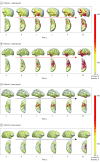
Figure 3.. Rapid Spread
Rapid spread vs slow spread as represented by fast β power activity mapped onto 3-dimensional reconstructions of 4 different patients’ brains (A-D). The rapid spread examples (A and B) show a spread to the lateral temporal cortex, including the posterior temporal lobe and superior temporal gyrus, in less than 10 seconds. A quantification of fast β Power showed that patients whose seizures recurred after surgery (A and B) had statistically significant increases in β power in the lateral temporal cortex outside standard anteromedial temporal resection margins during the first 10 seconds of ictus (P < .05) compared with patients who were seizure-free after surgery (mean, 137.5%; SEM, 16.8% vs 93.4%; SEM, 4.6%). β Power differences between these 2 groups in the orbitofrontal and extratemporal regions did not reach statistical significance. The numbers between the upper and lower frames represent seconds associated with seizure onset at a time of 0 seconds. The small dots on the brain images represent the position of intracranial electroencephalogram (EEG) electrodes. The pale yellow coloration (100% baseline) represents no change in β power compared with the mean baseline power. The areas without blue dots and without coloration have no iEEG electrode and thus no available power data. Slow spread examples show an intense foci of β power increases that is limited to mesial temporal structures and the temporal pole almost entirely within the margins of anteromedial temporal resection. aP < .05.
Figure 4.
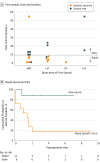
Figure 4.. Rapid Spread Outcomes
Intracranial electroencephalogram (iEEG) analyses. A, Time to first spread and location of first spread. Patients whose seizures recurred after surgery predominately spread initially either within the anteromedial temporal lobe (AMT) or to the ipsilateral temporal neocortex (LaT) before spreading to other regions. Patients whose seizures recurred showed significantly shorter latency to the first spread on visual iEEG analyses (P < .05). B, Patients whose seizures spread from their site of onset in less than 10 seconds (11 [55%]) were significantly more likely (hazard ratio, 5.99; 95% CI, 1.7-21.1; P < .01) to have recurrent seizures after anteromedial temporal resection than patients selected for iEEG whose seizures did not spread in less than 10 seconds (9 [45%]). ExT indicates extratemporal cortex; OF, orbitofrontal cortex.

References

    1. Wiebe S, Blume WT, Girvin JP, Eliasziw M; Effectiveness and Efficiency of Surgery for Temporal Lobe Epilepsy Study Group . A randomized, controlled trial of surgery for temporal-lobe epilepsy. N Engl J Med. 2001;345(5):311-318. doi:10.1056/NEJM200108023450501 - DOI - PubMed
    1. Dwivedi R, Ramanujam B, Chandra PS, et al. . Surgery for drug-resistant epilepsy in children. N Engl J Med. 2017;377(17):1639-1647. doi:10.1056/NEJMoa1615335 - DOI - PubMed
    1. Harroud A, Bouthillier A, Weil AG, Nguyen DK. Temporal lobe epilepsy surgery failures: a review. Epilepsy Res Treat. 2012;2012:201651. - PMC - PubMed
    1. Ryvlin P, Kahane P. The hidden causes of surgery-resistant temporal lobe epilepsy: extratemporal or temporal plus? Curr Opin Neurol. 2005;18(2):125-127. doi:10.1097/01.wco.0000162852.22026.6f - DOI - PubMed
    1. Barba C, Rheims S, Minotti L, et al. . Temporal plus epilepsy is a major determinant of temporal lobe surgery failures. Brain. 2016;139(pt 2):444-451. doi:10.1093/brain/awv372 - DOI - PubMed

Publication types