Review
. 2019 Apr 1;11(4):293-305.
doi: 10.1093/jmcb/mjy072.

Mutant p53 in cancer therapy-the barrier or the path

Affiliations
Review

Mutant p53 in cancer therapy-the barrier or the path

Xiang Zhou et al. J Mol Cell Biol. .

Abstract

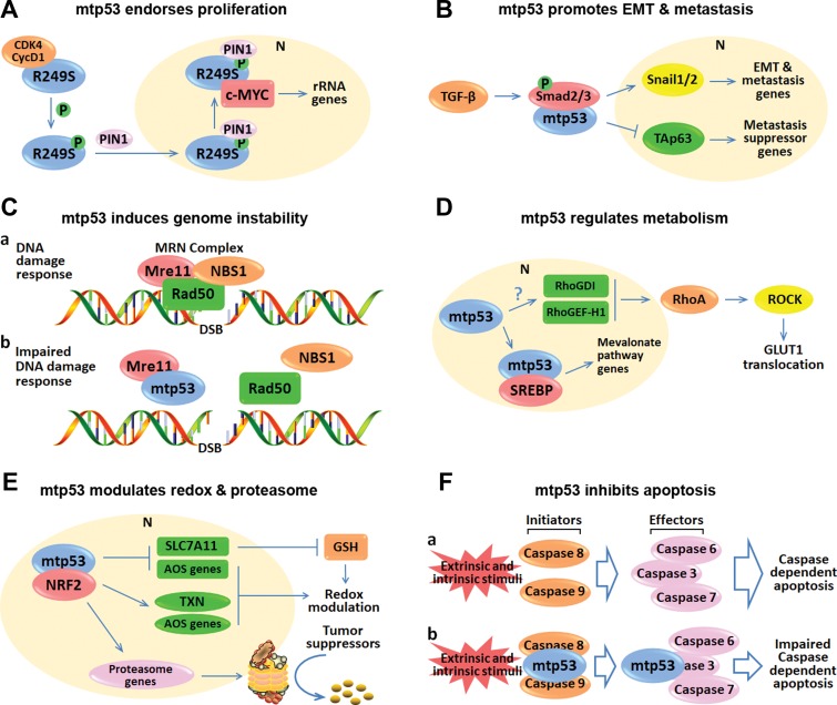
Since wild-type p53 is central for maintaining genomic stability and preventing oncogenesis, its coding gene TP53 is highly mutated in ~50% of human cancers, and its activity is almost abrogated in the rest of cancers. Approximately 80% of p53 mutations are single point mutations with several hotspot mutations. Besides loss of function and dominant-negative effect on the wild-type p53 activity, the hotspot p53 mutants also acquire new oncogenic functions, so-called 'gain-of-functions' (GOF). Because the GOF of mutant p53 is highly associated with late-stage malignance and drug resistance, these p53 mutants have become hot targets for developing novel cancer therapies. In this essay, we review some recent progresses in better understanding of the role of mutant p53 GOF in chemoresistance and the underlying mechanisms, and discuss the pros and cons of targeting mutant p53 for the development of anti-cancer therapies.

Keywords: cancer therapy; chemoresistance; gain-of-function; mutant p53; synthetic lethality.


Figures

Figure 1
Figure 1
p53 mutants promote cancer development and therapeutic resistance through their GOF.
Figure 2
Figure 2
Representative working modes of mutant p53 GOF. (A) p53-R249S is phosphorylated by CDK4/cyclin D1 and translocated into the nucleus as mediated by PIN1. The nuclear R249S binds to and augments c-Myc activity, leading to enhanced ribosome biogenesis and proliferation. (B) Mutant p53 prompts TGF-β-mediated EMT and metastasis by binding to Smad2/3. The interaction between mutant p53 and Smads leads to elevation of the downstream EMT and metastasis genes. Also, mutant p53, Smads, and TAp63 form a ternary complex that suppresses the anti-metastasis activity of TAp63. (C) Mutant p53 interferes with the assembly of the Mre11–Rad50–NBS1 complex on DNA double-stranded breaks, leading to genome instability. (D) Mutant p53 transcriptionally induces RhoGD1 and RhoGEF-H1 (Mizuarai et al., 2006; Bossi et al., 2008) through mechanisms to be identified, which elicits RhoA–ROCK–GLUT1 cascade, leading to increased aerobic glycolysis. Additionally, mutant p53 binds to the transcription factor SREBP, leading to the activated mevalonate pathway responsible for the regulation of lipid metabolism. (E) Mutant p53 modulates redox and proteasome by interacting with NRF2. Mutant p53 can induce and repress NRF2 target genes to control the glutathione (GSH) level and the redox balance. Moreover, mutant p53 promotes the expression of NRF2 target genes involved in proteasome machinery, leading to increased proteasomal degradation of tumor suppressors. (F) Mutant p53 suppresses apoptosis by directly binding to caspases. Upon external and/or internal stimuli, the initiator caspases, caspase-8, and caspase-9, can proteolytically activate the effector caspases, caspase-3, caspase-6, and caspase-7, thus triggering the caspase-dependent apoptosis. However, mutant p53 can interact with and inhibit caspase-8, caspase-9, and caspase-3 to compromise the caspase-dependent apoptosis. In conclusion, p53 mutants execute their oncogenic GOF by interacting with transcription factors or cofactors to promote (A, B, D, E) or repress (B, E) gene expression, or with proteins irrelevant to transcription (C, F). mtp53 indicates mutant p53.
Figure 3
Figure 3
Therapeutic strategies targeting mutant p53. (A) Ablation of mutant p53 in cancer is one of the most effective strategies for developing cancer therapy. HSP90 is highly expressed in cancer and crucial to mutant p53 stabilization. HSP90 inhibitors block HSP90 function and lead to MDM2- or CHIP-mediated proteasomal degradation of mutant p53 proteins. In addition, both Spautin-1 and MCB-613 can induce lysosomal degradation of mutant p53 through HSPA8/LAMP2A-mediated CMA pathway and inhibition of the deubiquitinase USP15, respectively. Moreover, HDAC inhibitors can suppress mutant p53 gene transcription. (B) Another attractive strategy is to restore the mutant p53 to a wild-type-resembling conformation. APR-246, COTI-2, and PK11007 can reactivate mutant p53 and trigger the expression of wild-type p53 target genes. Also, APR-246 and PK11007 can kill cancer cells by elevating the ROS level and inducing oxidative stress. (C) The TP53 gene mutations also impose vulnerability on cancer cells. In response to DNA damage stress, wild-type p53-mediated G1/S arrest and WEE1-mediated G2/M arrest are essential for cells to repair damaged DNA. Nevertheless, the WEE1-mediated G2/M checkpoint renders crucial survival dependency in the mutant p53-sustaining cancer cells with defective G1/S checkpoint. Thus, the WEE1 inhibitor can cause synthetic lethality to these cancer cells by abrogating the remaining G2/M checkpoint. mtp53 indicates mutant p53.

References

    1. Aas T., Borresen A.L., Geisler S., et al. . (1996). Specific P53 mutations are associated with de novo resistance to doxorubicin in breast cancer patients. Nat. Med. 2, 811–814. - PubMed
    1. Adorno M., Cordenonsi M., Montagner M., et al. . (2009). A mutant-p53/Smad complex opposes p63 to empower TGFβ-induced metastasis. Cell 137, 87–98. - PubMed
    1. Agarwal S., Bell C.M., Taylor S.M., et al. . (2016). p53 deletion or hotspot mutations enhance mTORC1 activity by altering lysosomal dynamics of TSC2 and Rheb. Mol. Cancer Res. 14, 66–77. - PMC - PubMed
    1. Aggarwal M., Saxena R., Sinclair E., et al. . (2016). Reactivation of mutant p53 by a dietary-related compound phenethyl isothiocyanate inhibits tumor growth. Cell Death Differ. 23, 1615–1627. - PMC - PubMed
    1. Alam S.K., Yadav V.K., Bajaj S., et al. . (2016). DNA damage-induced ephrin-B2 reverse signaling promotes chemoresistance and drives EMT in colorectal carcinoma harboring mutant p53. Cell Death Differ. 23, 707–722. - PMC - PubMed

Publication types

MeSH terms

Substances