. 2019 Jun;95(6):664-671.
doi: 10.1002/cyto.a.23642. Epub 2018 Dec 3.

Detection of Apoptotic Circulating Tumor Cells Using in vivo Fluorescence Flow Cytometry

Affiliations

Detection of Apoptotic Circulating Tumor Cells Using in vivo Fluorescence Flow Cytometry

Jacqueline Nolan et al. Cytometry A. 2019 Jun.

Abstract

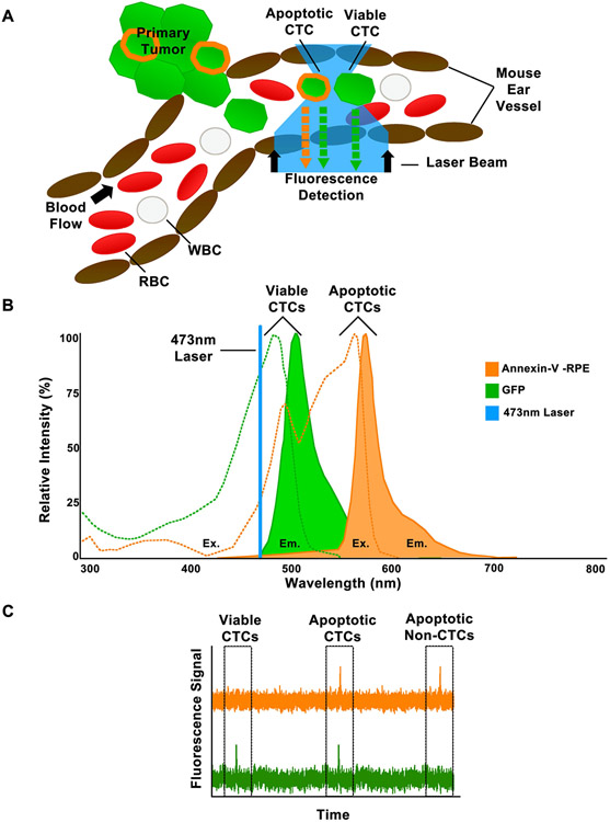
Most cancer patients die from metastatic disease as a result of a circulating tumor cell (CTC) spreading from a primary tumor through the blood circulation to distant organs. Many studies have demonstrated the tremendous potential of using CTC counts as prognostic markers of metastatic development and therapeutic efficacy. However, it is only the viable CTCs capable of surviving in the blood circulation that can create distant metastasis. To date, little progress has been made in understanding what proportion of CTCs is viable and what proportion is in an apoptotic state. Here, we introduce a novel approach toward in situ characterization of CTC apoptosis status using a multicolor in vivo flow cytometry platform with fluorescent detection for the real-time identification and enumeration of such cells directly in blood flow. The proof of concept was demonstrated with two-color fluorescence flow cytometry (FFC) using breast cancer cells MDA-MB-231 expressing green fluorescein protein (GFP), staurosporine as an activator of apoptosis, Annexin-V apoptotic kit with orange dye color, and a mouse model. The future application of this new platform for real-time monitoring of antitumor drug efficiency is discussed. © 2018 International Society for Advancement of Cytometry.

Keywords: apoptosis; circulating tumor cells; fluorescence flow cytometry; in vivo detection of circulating tumor cells.


Figures

Figure 1.
Figure 1.
In vivo Fluorescence Flow Cytometry (FFC) Diagnostic Platform Schematic. A. The in vivo schematic for detection of viable and apoptotic CTCs using FFC. B. Excitation and Emission spectrum of GFP (CTC label) and Annexin-V-PRE (apoptotic CTC label). C. The expected fluorescence signal from the photodetector for viable CTCs, apoptotic CTCs, and apoptotic non-CTCs.
Figure 2.
Figure 2.
In Vitro Cell Viability Testing of Cells Treated With Staurosporine. Two assays were used to determine cell viability of MDA-GFP cells after the treatment of 25 μg/mL of Staurosporine. Fluorescence microscopy (A) was performed using Annexin-V-RPE to visualize cells undergoing apoptosis. The trypan blue exclusion assay (B) was used to determine the overall viability of treated cells compared to untreated control cells at 3 time points (1,3, and 24 hours).
Figure 3.
Figure 3.
In Vitro Fluorescent Flow Cytometry (FFC) detection of apoptotic mimic CTCs. Using a 50 μM capillary tube was performed on control MDA-GFP cells (A) and Staurosporine treated MDA-GFP cells (B). Each group was incubated with Annexin-V-RPE (orange) before FFC was performed. On the left, fragments of each FFC trace are shown with the green fluorescence trace on top and the orange fluorescence trace on the bottom. 2D analysis of each trace is represented on the right with orange fluorescence on the y-axis and green fluorescence on the x-axis.
Figure 4.
Figure 4.
Monitoring of Ex Vivo labeled MDA-GFP cells using In Vivo Fluorescence Flow Cytometry (FFC). Control MDA-GFP cells (A) and MDA-GFP cells treated with Staurosporine (B) for 30 min were labeled with annexin-V-RPE (orange) Ex Vivo and then injected (i.v) into a mouse. In Vivo FFC was used to monitor a vessel in the ear of the mouse.
Figure 5.
Figure 5.
In Vivo detection of apoptotic circulating blood and tumor cells using FFC. A 50-μm mouse ear vessel was monitored using our in vivo FFC platform. Annexin-V-RPE (A) or Annexin-V-RPE and 25 μg/mL of starurosporine (B) was injected intravenously (i.v.) and orange fluorescent signal was observed. Then Annexin-V-RPE and MDA-GFP cells that had previously been incubated with 25 μg/mL of staurosporine (C) were intravenously injected into the mouse. Green (top) and orange (bottom) fluorescent signal was observed. The inset shows a coincidental peak that occurred in both the orange channel and the green channel.

References

    1. Fidler IJ. The pathogenesis of cancer metastasis: the ‘seed and soil’ hypothesis revisited. Nat Rev Cancer. 2003;3(6):453–8. - PubMed
    1. Massague J, Obenauf AC. Metastatic colonization by circulating tumour cells. Nature. 2016;529(7586):298–306 - PMC - PubMed
    1. Nguyen DX, Bos PD, Massague J. Metastasis: from dissemination to organ-specific colonization. Nature Reviews Cancer. 2009;9(4):274–284. - PubMed
    1. Kim M, Oskarsson T, Acharyya S, Nguyen DX, Zhang X, Norton L, Massague J. Tumor self-seeding by circulating cancer cells. Cell. 2009;139(7):1315–1326. - PMC - PubMed
    1. Talmadge JE, Fidler IJ. AACR centennial series: the biology of cancer metastasis: historical perspective. Cancer Research. 2010;70(14):5649–5669. - PMC - PubMed

Publication types

MeSH terms