. 2019 Jan:20:526-532.
doi: 10.1016/j.redox.2018.11.011. Epub 2018 Nov 22.

Proteomic analysis of microbial induced redox-dependent intestinal signaling

Affiliations

Proteomic analysis of microbial induced redox-dependent intestinal signaling

Jason D Matthews et al. Redox Biol. 2019 Jan.

Abstract

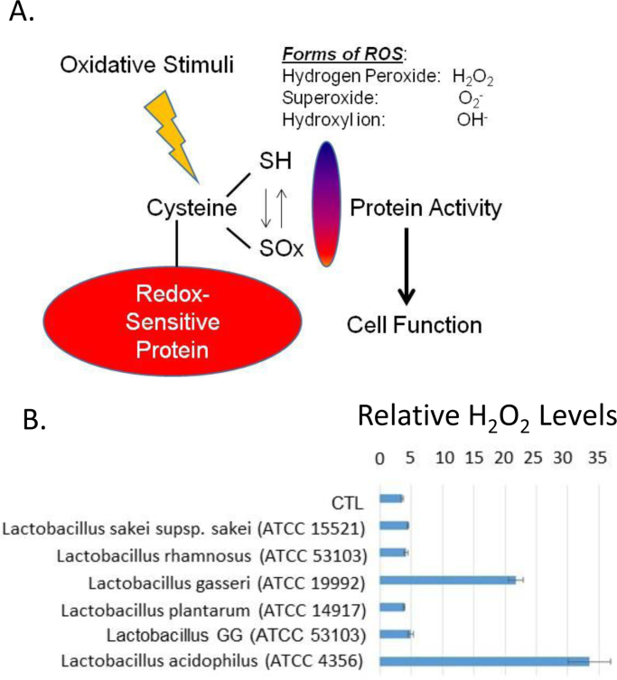
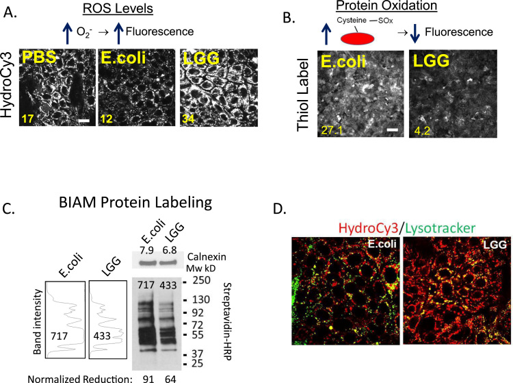
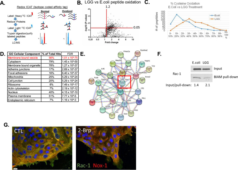
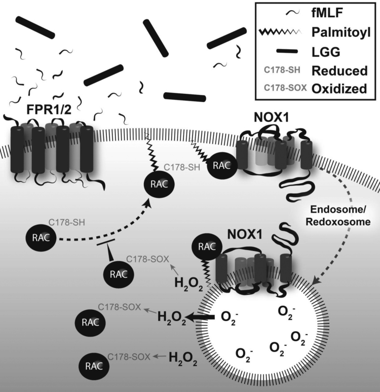
Intestinal homeostasis is regulated in-part by reactive oxygen species (ROS) that are generated in the colonic mucosa following contact with certain lactobacilli. Mechanistically, ROS can modulate protein function through the oxidation of cysteine residues within proteins. Recent advances in cysteine labeling by the Isotope Coded Affinity Tags (ICATs) technique has facilitated the identification of cysteine thiol modifications in response to stimuli. Here, we used ICATs to map the redox protein network oxidized upon initial contact of the colonic mucosa with Lactobacillus rhamnosus GG (LGG). We detected significant LGG-specific redox changes in over 450 proteins, many of which are implicated to function in cellular processes such as endosomal trafficking, epithelial cell junctions, barrier integrity, and cytoskeleton maintenance and formation. We particularly noted the LGG-specific oxidation of Rac1, which is a pleiotropic regulator of many cellular processes. Together, these data reveal new insights into lactobacilli-induced and redox-dependent networks involved in intestinal homeostasis.


Figures

Fig. 1
Fig. 1
Cellular or bacterial derived ROS can alter protein function. (A) Schematic of cysteine oxidation to regulate cell function. (B) Various strains of Lactobacillus were tested for their ability to produce hydrogen peroxide. Bacteria were grown in overnight cultures and the supernatants tested against a negative PBS control for the relative abundance of hydrogen peroxide in a 96-well plate using a ROS-GLO kit. A luminometer was used to quantify the levels of hydrogen peroxide.
Fig. 2
Fig. 2
LGG induces ROS that oxidizes cysteines. (A) Intestinal epithelial cells (SKCO15) were contacted with 108 CFU/mL of E. coli or LGG for 15 min, and then with 15 µM HydroCy3 for 30 min before confocal microscopic analysis at 555 nm, (scale bars 20 µm). (B) SKCO15 were contacted with 108 CFU/mL of E. coli or LGG for 15 min, labeled for 30 min with a thiol-reactive, Thiol Tracker™ fluorescence probe, and then analyzed by fluorescence microscopy at 405 nm (scale bars 200 µm). Mean image intensity is shown at bottom left for A and B. (C) Biotinylated-iodoacetamide (BIAM) labeling of cysteine residues in lysates of LGG or E. coli contacted SKCO15 cells, followed by pull-down of labeled residues with streptavidin conjugated agarose and detected by Western blot using HRP conjugated streptavidin as a probe. The relative intensity of each lane of the blot is shown in arbitrary units to the left. Each value was normalized to calnexin that served as a loading control to give the relative oxidation amounts. (D) Dual labeling of LGG or E. coli contacted (15 mins) SKCO15 cultured cells with HydroCy3 (red) and Lysotracker (green). Note co-localization of lysotracker and hydro-Cy3 in LGG contacted cells (bars 10 µm).
Fig. 3
Fig. 3
ICAT analysis of intestinal epithelial scrapings from Germ-free mice given a rectal lavage containing 108 CFU/mL of LGG or E. coli for 15 min (A) Graphical representation of ICAT labeling procedure. (B) Volcano plot of peptide hits (~1500) differentially oxidized in LGG contacted colon compared to E.coli contacted colon. (C) Relative peptide oxidation levels significantly changed in LGG or E.coli contacted colon. (D) Pathways analysis of peptide hits significantly oxidized in LGG samples (hits from upper right quadrant in A). (E) STRING map of proteins involved in membrane bound vesicle pathway, including Rac1. (F) BIAM labeling of cell lysates from SKCO15 cells after 15 min contact with 108 CFU/mL of LGG or E. coli for 15 min. Inputs lysates, and pull-down lysates were analyzed by immunoblot using an antibody against Rac1. (G) SKCO15 cells exhibit decreased subcellular co-localization between Rac1 (green) and Nox1 (yellow) after exposed to 25 µM 2-Brp for 2 h, (scale bar µm).
Fig. 4
Fig. 4
Putative model for LGG regulation of ROS production in the gut epithelium. Rac1 is first activated following LGG stimulation of Formyl Peptide Receptor. Rac1 subsequently becomes palmitoylated at C178 and localized at the plasma membrane, where it activates Nox-1 to generate ROS. Endocytosis of this complex forms a redoxosome that generates elevated levels of ROS. In a feedback loop, further palmitoylation of Rac-1 is blocked by oxidation of C178, decreasing the formation of redoxosomes.

References

    1. Abt M.C., Mckenney P.T., Pamer E.G. Clostridium difficile colitis: pathogenesis and host defence. Nat. Rev. Microbiol. 2016;14:609–620. - PMC - PubMed
    1. Alam A., Leoni G., Quiros M., Wu H., Desai C., Nishio H., Jones R.M., Nusrat A., Neish A.S. The microenvironment of injured murine gut elicits a local pro-restitutive microbiota. Nat. Microbiol. 2016;1:15021. - PMC - PubMed
    1. Alam A., Leoni G., Wentworth C.C., Kwal J.M., Wu H., Ardita C.S., Swanson P.A., Lambeth J.D., Jones R.M., Nusrat A., Neish A.S. Redox signaling regulates commensal-mediated mucosal homeostasis and restitution and requires formyl peptide receptor 1. Mucosal Immunol. 2014;7:645–655. - PMC - PubMed
    1. Babbin B.A., Jesaitis A.J., Ivanov A.I., Kelly D., Laukoetter M., Nava P., Parkos C.A., Nusrat A. Formyl peptide receptor-1 activation enhances intestinal epithelial cell restitution through phosphatidylinositol 3-kinase-dependent activation of Rac1 and Cdc42. J. Immunol. 2007;179:8112–8121. - PubMed
    1. Chen R.C., Xu L.M., Du S.J., Huang S.S., Wu H., Dong J.J., Huang J.R., Wang X.D., Feng W.K., Chen Y.P. Lactobacillus rhamnosus GG supernatant promotes intestinal barrier function, balances Treg and TH17 cells and ameliorates hepatic injury in a mouse model of chronic-binge alcohol feeding. Toxicol. Lett. 2016;241:103–110. - PubMed

Publication types

LinkOut - more resources